
Page 1322 - Williams Hematology ( PDFDrive )
P. 1322
1296 Part X: Malignant Myeloid Diseases Chapter 84: Polycythemia Vera 1297
DIFFERENTIAL DIAGNOSIS out congenital polycythemia should always be undertaken in patients
with atypical presentations; however, the presence of polycythemia in
Also refer to Chap. 34, Table 34–2, and Chap. 57, Fig. 57–6. other relatives does not rule out PV.
PV has to be differentiated from spurious polycythemia, second-
ary polycythemias, congenital disorders of hypoxia sensing, and pri-
mary congenital polycythemias. The differential diagnostic task has TREATMENT
been facilitated by the discovery of the JAK2 V617F mutation that is pres- The major causes of morbidity and mortality in PV are an increased
ent in 95 percent or more of all PV patients. 165,166 Thus, the majority of incidence of vascular complications (i.e., thrombosis and/or hemor-
PV patients can be diagnosed with a complete blood count repeated rhage), and progression to MF or acute leukemia/myelodysplasia. In the
twice and molecular studies to confirm the presence of the JAK2 V617F first randomized trial of PV patients, a history of previous thrombosis,
mutation. age, treatment with phlebotomies, and rate of phlebotomies contributed
For the remainder of polycythemic patients, additional diagnostic to the increased risk of thrombosis. Presently, the age of the patient
76
measures need to be undertaken. These may include measuring serum (>60 years) and previous thrombotic events are universally acknowl-
erythropoietin level, measuring venous oxygen saturation to calcu- edged major risk factors for major vascular complications in PV. 71
late hemoglobin P50, measuring arterial oxygen saturation (less than Thus, PV patients are classified as low risk or high risk, with age
92 percent suggests cardiac or pulmonary etiologies), abdominal CT greater than 60 years and previous thrombotic events (including tran-
scan (to exclude renal, hepatic, and cerebral tumors), brain magnetic sient ischemic attacks) defining the high-risk category. The assigned risk
resonance imaging (to rule out a cerebellar hemangioblastoma), and classification has a major impact on therapeutic decisions, as high-risk
detailed family studies. patients are treated with cytoreductive therapies. Other risk factors may
If a diagnosis at this point has not yet been made, the patient could also play a role in the pathogenesis of thrombosis, such as hypertension,
be referred to a specialized center for further testing. These studies may diabetes, or smoking, as well as leukocytosis 172–176 and JAK2 V617F muta-
171
include measuring changes in serum erythropoietin levels after phle- tional allele burden. 80,177 There is a need for prospective clinical studies
botomies, red blood cell and plasma volume studies (to diagnose spuri- with stratification of patients according to these criteria, but until such
ous polycythemia or masked PV), genomic sequencing studies, testing evidence is available, patients with high leukocyte levels and/or high
for JAK2 exon 12 mutations, and in vitro studies for EECs. The latter JAK2 V617F mutational allele burden should be managed according to
two are used to diagnose JAK2 V617F -negative PV patients (<5 percent of conventional criteria.
all PVs). An elevated platelet count does not increase the risk of thrombosis,
Distinguishing between PV and other polycythemic disorders but it may increase the risk of hemorrhage. Bleeding is more frequent
128
may, at times, be challenging. Although the diagnosis of PV may be in patients with platelet counts in excess of 1500 × 10 /L, thought to be
9
straightforward if patients have the classic features of PV as defined the result of an acquired type 2 von Willebrand disease.
by the most recent WHO guidelines, patients often present with an Treatment of PV depends on risk evaluation at diagnosis and evo-
154
incomplete phenotype. Some of the clinical and laboratory features lution of the disease over time. Treatment should be given both to alle-
that can be helpful for differential diagnosis are summarized in Chap. viate symptoms and prevent complications. Updated consensus-based
34, Table 34–2 and Chap. 57, Fig. 57–6. While the current WHO diag- guidelines for the management of PV and other major MPNs were pub-
nostic criteria (presented in Table 84–1) represent an improvement lished by a panel of experts formed by European LeukemiaNet (ELN).
154
178
over previous guidelines, they do not necessarily discriminate between Response criteria by which new therapies are evaluated were also
individual MPNs, and are still a matter of some debate. 168,169 Children updated to facilitate direct comparison of therapeutic efficacy across
167
with PV are especially unlikely to fit the most recent WHO criteria. clinical trials (Table 84–2). This joint effort between ELN and the Inter-
170
179
Most importantly, often laborious and time-consuming efforts to rule
national Working Group for Myeloproliferative Neoplasms Research
and Treatment (IWG-MRT) provides updated guidelines incorporating
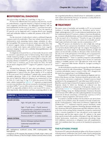
TABLE 84–1. World Health Organization Criteria for the assessment of clinical, hematologic, and histologic response, as well as
Diagnosis of Polycythemia Vera, 2008 symptoms, disease progression, and vascular events. 179
It is useful to consider treatment for the plethoric and spent phases
MAJOR CRITERIA separately. In the plethoric phase, the mainstay of therapy remains non-
1 Hgb >18.5 g/dL (men), >16.5 g/dL (women) specific myelosuppression, which many practitioners supplement with
or phlebotomies. 82,178,180 Additional measures prevent thrombotic events
Hgb >17 g/dL (men), >15 g/dL (women) (i.e., aspirin) and relieve symptoms. Promising therapies include pegy-
lated interferon (PEG-IFN) preparations and JAK2 inhibitors; these are,
if associated with a sustained increase of ≥2 at the time of this writing, being evaluated by prospective randomized
g/dL from baseline that cannot be attributed trials. A minority of patients (i.e., low-risk patients) can be treated with
to correction of iron deficiency
phlebotomies and low-dose aspirin alone. PV patients in the spent
2 Presence of JAK2 V617F or similar mutation phase may be treated with a number of therapies (Chap. 86), which may
MINOR CRITERIA include hydroxyurea, transfusion, erythropoiesis-stimulating drugs,
JAK2 inhibitors, splenectomy, or allogeneic stem cell transplantation;
1 Marrow trilineage myeloproliferation
only JAK2 inhibitors have been proven to be beneficial in prospective
2 Subnormal serum EPO level trials. 235
3 EEC growth
THE PLETHORIC PHASE
EEC, endogenous erythroid colony; EPO, erythropoietin; Hgb,
hemoglobin. Treatment of PV patients in the plethoric phase of the disease is aimed at
A diagnosis of PV requires the presence of both major criteria and reducing marrow proliferation and blood counts, thereby ameliorating
one minor criterion, or the first major criterion and two minor criteria. symptoms and decreasing the risk of thrombosis and bleeding. 180,181 This
Kaushansky_chapter 84_p1291-1306.indd 1297 9/21/15 11:11 AM