
Page 1555 - Williams Hematology ( PDFDrive )
P. 1555
1530 Part XI: Malignant Lymphoid Diseases Chapter 92: Chronic Lymphocytic Leukemia 1531
CLINICAL PRESENTATION OF CHRONIC
LYMPHOCYTIC LEUKEMIA
CLL is a disease of the elderly with the median age at diagnosis being 72
years. Most patients are asymptomatic at diagnosis and are diagnosed
as a result of incidental finding of lymphadenopathy and lymphocytosis
of uncertain etiology as part of an evaluation unrelated to CLL. A vast
majority of patients may not have any significant symptoms related to
the disease but some patients may experience mild fatigue or minor lim-
itations in their activities of daily living. A subset of patients may present
with recurring infectious complications, especially upper respiratory
tract infections. Patients with advanced disease can uncommonly pres-
ent with drenching night sweats, fevers, and weight loss (B symptoms),
and signs and symptoms related to anemia, thrombocytopenia, and
lymphadenopathy. The lymphadenopathy typically observed in patients
with CLL is generally not fixed or tender and very rarely causes symp-
toms of organ dysfunction or resulting dependent-limb lymphedema.
Patients can have exacerbations of their lymphadenopathy during an Figure 92–1. Typical chronic lymphocytic leukemia blood film. (Repro-
acute infectious episode, but this typically returns to baseline upon duced with permission from Ash Image Bank, Peter Maslak, 2013, © the
resolution of the underlying infectious complication. Splenomegaly American Society of Hematology.)
99
is seen commonly in patients with CLL with resultant hypersplenism
and thrombocytopenia. Significant hepatomegaly because of leukemic
infiltration is unusual. CLL infiltration of multiple organs has been film, an albumin preparation is sometimes required. Patients can also
described but these are typically seen in patients with advanced disease have large prolymphocytes with prominent nucleoli in the blood but
and will occasionally cause symptoms. Pulmonary involvement has these lymphocytes must be less than 55 percent of the total lymphocyte
been observed in patients with high lymphocyte count and typically population. The anemia is typically normocytic and normochromic
presents as an interstitial infiltrate on chest radiography. Chylous and and platelet morphology is typically preserved. A marrow aspirate and
hemorrhagic pleural effusions have also been reported. 100–102 Similarly, biopsy is not required for the vast majority of patients with CLL at ini-
leukemic infiltration of the gastrointestinal tract may result in chronic tial presentation to establish a diagnosis. However, we do recommend
diarrhea or iron-deficiency anemia secondary to chronic bleeding or performing a marrow aspirate and biopsy in patients with anemia and
malabsorption. However, this mucosal infiltration is more commonly thrombocytopenia to evaluate the presence of autoimmune hemolytic
seen in patients with mantle cell lymphoma. CLL involvement of the anemia and/or immune thrombocytopenia. Marrow biopsy typically
central nervous system is rare and may result in headaches, confu- shows diffuse marrow involvement with a monotypic population of
sion, meningismus, or cranial nerve palsies. More commonly, these small lymphocytes. Variability of marrow involvement has been histor-
103
patients are at higher risk for opportunistic infections of the central ically used as a potential marker for prognosis but has limited appli-
nervous system because of their deficient immune system. Patients with cability given the availability of more specific and sensitive prognostic
CLL are also known to have insect bite hypersensitivity. 104,105 Patient’s markers. 106,107 The red cell precursors and megakaryocytes usually dis-
typically present with recurrent, erythematous, painful eruptions usu- play an unremarkable morphology but do diminish in numbers with
ally on the exposed part of the extremities. Evaluation of skin biopsies
from these patients reveal a mixed infiltrating population of T cells, B
cells, and eosinophils. These resolve over time and can be effectively
treated with a short course of glucocorticoid.
EVALUATION OF THE PATIENT WITH
CHRONIC LYMPHOCYTIC LEUKEMIA
According to the IWCLL-2008 criteria, the diagnosis of CLL requires
a sustained monoclonal lymphocytosis of greater than 5000 cells/μL of
monoclonal B cells. This requires blood flow cytometry for immuno-
1
phenotyping the B cells, which additionally reveals the cells to be pos-
itive for CD19, dim CD20, dim surface immunoglobulin, and negative
for CD10, CD79b, and FMC7. A similar disease presentation with no
evidence of hematopoietic involvement and with only lymph node
involvement by cells of comparable morphology will be classified as
small lymphocytic lymphoma. These patients are essentially managed
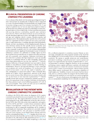
similar to patients with CLL. CLL cells appear as small blue lympho-
cytes with scant cytoplasm on the Wright-Giemsa staining commonly
used for evaluating blood films (Fig. 92–1). Smudge cells are also com-
monly observed on the blood film and this results from the mechanical Figure 92–2. Typical chronic lymphocytic leukemia blood film with
disruption of the cells during the slide preparation process (Fig. 92–2). smudge cells. (Reproduced with permission from ASH image bank, Peter
For an improved evaluation of the cellular morphology on the blood Maslak, 2010. © the American Society of Hematology.)
Kaushansky_chapter 92_p1527-1552.indd 1530 9/18/15 10:47 AM