
Page 2074 - Williams Hematology ( PDFDrive )
P. 2074
2048 Part XII: Hemostasis and Thrombosis Chapter 120: Hereditary Qualitative Platelet Disorders 2049
N in the remaining GPIbβ allele have included P96S and P29L. 236,237 In
other studies of patients with the 22q11.2 deletion syndrome, modest
reductions in platelet count and increases in platelet volume, as well as
reduced platelet agglutination to ristocetin and decreased platelet GPIb/
IX expression, have been variably reported, consistent with hemizygos-
Bernard-Soulier ity for GPIbβ. 240–244
Leucine- syndrome mutations A number of monoallelic, heterozygous mutations in the genes
rich of the GPIb–IX complex have been described as causing macrothrom-
repeats bocytopenia, some, but not all, of which have also been implicated in
causing the biallelic form either because of homozygosity or compound
Platelet-type heterozygosity. These include a heterozygous mutation in the second
154
VWD
245
mutations leucine-rich repeat (L57F) in which the affected patients have mod-
erate bleeding symptoms, moderate thrombocytopenia, and giant plate-
Regulatory lets. Additional monoallelic mutations of GPIbα that appear to produce
loop dominant effects are N41H and Y54D. 247
246
The “Bolzano” defect, which involves a mutation in the sixth
leucine-rich repeat of GPIbα (A156V), results in a GPIbα molecule that
has reduced ability to bind VWF, but can bind thrombin. It has been
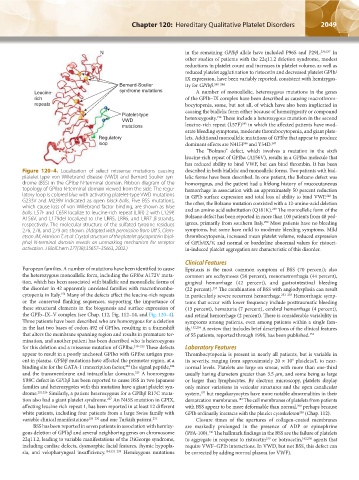
Figure 120–4. Localization of select missense mutations causing described in both biallelic and monoallelic forms. Two patients with bial-
platelet-type von Willebrand disease (VWD) and Bernard-Soulier syn- lelic forms have been described. In one patient, the Bolzano defect was
drome (BSS) in the GPIbα N-terminal domain. Ribbon diagram of the homozygous, and the patient had a lifelong history of mucocutaneous
topology of GPIbα N-terminal domain viewed from the side. The regu- hemorrhage in association with an approximately 50 percent reduction
latory loop is colored blue with activating platelet-type VWD mutations in GPIb surface expression and total loss of ability to bind VWF. In
248
G233V and M239V indicated as open black balls. Five BSS mutations, the other, the Bolzano mutation coexisted with a 12-amino-acid deletion
which cause loss of von Willebrand factor binding, are shown as blue and an amino acid substitution (Q181K). The monoallelic form of the
249
balls. L57F and C65R localize to leucine-rich repeat (LRR) 2 with L129P,
A156V, and L179del localized to the LRR5, LRR6, and LRR7 β-strands, Bolzano defect has been reported in more than 100 patients from 48 ped-
250
respectively. The molecular structure of the sulfated tyrosine residues igrees, primarily from southern Italy. Most patients have no bleeding
276, 278, and 279 are shown. (Adapted with permission from Uff S, Clem- symptoms, but some have mild to moderate bleeding symptoms. Mild
etson JM, Harrison T, et al: Crystal structure of the platelet glycoprotein Ib(al- thrombocytopenia, increased mean platelet volume, reduced expression
pha) N-terminal domain reveals an unmasking mechanism for receptor of GPIb/IX/V, and normal or borderline abnormal values for ristocet-
activation. J BiolChem 277(38):35657–35663, 2002.) in-induced platelet aggregation are characteristic of this disorder.
Clinical Features
European families. A number of mutations have been identified to cause Epistaxis is the most common symptom of BSS (70 percent); also
the heterozygous monoallelic form, including the GPIbα A172V muta- common are ecchymoses (58 percent), menometrorrhagia (44 percent),
tion, which has been associated with biallelic and monoallelic forms of gingival hemorrhage (42 percent), and gastrointestinal bleeding
the disorder in 42 apparently unrelated families with macrothrombo- (22 percent). The combination of BSS with angiodysplasia can result
157
cytopenia in Italy. Many of the defects affect the leucine-rich repeats in particularly severe recurrent hemorrhage. 251–253 Hemorrhagic symp-
154
or the conserved flanking sequences, supporting the importance of toms that occur with lower frequency include posttraumatic bleeding
these structural elements in the biogenesis and surface expression of (13 percent), hematuria (7 percent), cerebral hemorrhage (4 percent),
the GPIb–IX–V complex (see Chap. 112, Fig. 112–14, and Fig. 120–4). and retinal hemorrhage (2 percent). There is considerable variability in
Three patients have been described who are homozygous for a deletion symptoms among patients, even among patients within a single fam-
in the last two bases of codon 492 of GPIbα, resulting in a frameshift ily. 152,254 A review that includes brief descriptions of the clinical features
that alters the membrane spanning region and results in premature ter- of 55 patients, reported through 1998, has been published. 152
mination, and another patient has been described who is heterozygous
for this deletion and a missense mutation of GPIbα. 219–222 These defects Laboratory Features
appear to result in a poorly anchored GPIbα with GPIbα antigen pres- Thrombocytopenia is present in nearly all patients, but is variable in
ent in plasma. GPIbβ mutations have affected the promoter region, at a its severity, ranging from approximately 20 × 10 platelets/L to near-
9
binding site for the GATA-1 transcription factor, the signal peptide, normal levels. Platelets are large on smear, with more than one-third
223
224
and the transmembrane and intracellular domains. A homozygous usually having diameters greater than 3.5 μm, and some being as large
225
Y88C defect in GPIbβ has been reported to cause BSS in two Japanese or larger than lymphocytes. By electron microscopy, platelets display
families and heterozygotes with this mutation have a giant platelet syn- only minor variations in vesicular structures and the open canalicular
drome. 221,226 Similarly, a patient heterozygous for a GPIbβ R17C muta- system, but megakaryocytes have more notable abnormalities in their
157
tion also had a giant platelet syndrome. An N45S mutation in GPIX, demarcation membranes. The cell membranes of platelets from patients
227
182
affecting leucine rich repeat 1, has been reported in at least 12 different with BSS appear to be more deformable than normal, perhaps because
208
white patients, including four patients from a large Swiss family with GPIb ordinarily interacts with the platelet cytoskeleton (Chap. 112).
255
variable clinical manifestations 228–230 and one Turkish patient. 231 Closure times of the apertures of collagen-coated membranes
BSS has been reported in seven patients in association with hemizy- are markedly prolonged in the presence of ADP or epinephrine
gous deletion of GPIbβ and several neighboring genes on chromosome (PFA-100). The hallmark findings in the BSS are the failure of platelets
136
22q11.2, leading to variable manifestations of the DiGeorge syndrome, to aggregate in response to ristocetin or botrocetin, 162,256 agents that
159
including cardiac defects, dysmorphic facial features, thymic hypopla- require VWF–GPIb interactions. In VWD, but not BSS, this defect can
sia, and velopharyngeal insufficiency. 154,232–239 Hemizygous mutations be corrected by adding normal plasma (or VWF).
Kaushansky_chapter 120_p2039-2072.indd 2049 9/21/15 2:20 PM