
Page 2069 - Williams Hematology ( PDFDrive )
P. 2069
2044 Part XII: Hemostasis and Thrombosis Chapter 120: Hereditary Qualitative Platelet Disorders 2045
not occur normally, the very small amount of residual integrin α will Of note, many of the patients with identified mutations are compound
IIb
53
be pro-α , not mature α . Pro-α has been reported to bind to the heterozygotes rather than homozygotes, indicating that a sizable num-
IIb
IIb
IIb
membrane-bound endoplasmic reticulum chaperone calnexin, provid- ber of silent carriers are present in the population. Where consanguinity
ing a potential mechanism for assessing whether the protein has under- is common, the disorder is more likely to be caused by a homozygous
gone proper folding (calnexin cycle) and perhaps explaining how the mutation arising in a founder, but even under these circumstances, more
receptor adopts a bent configuration. 54,55 than one mutation may be present. Thus, in the Iraqi-Jewish population,
Integrin β (GPIIIa) can also combine with the integrin α in which consanguinity has been present from 586 bce to the present,
3
V
39
(CD51) subunit to form the integrin α β “vitronectin” receptor 30,56,57 two separate mutations have been identified in more than one family.
V 3
(see Fig. 120–2; Chap. 112). This receptor can bind many of the same Most of the missense mutations result in decreased expression of integ-
adhesive glycoproteins as integrin α β , although there are some dif- rin α β on the surface of platelets. This probably reflects the stringent
IIb 3
IIb 3
ferences in ligand preference and binding sequences. 57–61 A small num- structural requirements for proper folding and complex formation.
ber of integrin α β receptors are present on platelets (50 to 100 per Mutations in Integrin α β Within the Metal Ion-Dependent
IIb 3
V 3
platelet) 60,62,63 ; osteoclasts, endothelial cells, macrophages, vascular Adhesion Site of Integrin β and the Interface with the Integrin α
IIb
3
smooth muscle, and uterine cells, among others, also have integrin α β β-Propeller A metal coordination site or MIDAS domain, which is
V 3
receptors. 64,65 In general, GT patients with defects in integrin β also highly conserved in six integrin receptor α-chain subunits and required
3
are deficient in integrin α β , whereas patients with defects in integrin for ligand-binding, is also present in the β-A (or I-like) domain of the
V 3
α have either normal or increased numbers of platelet integrin α β integrin β subunit. Mutagenesis and molecular modeling experi-
72
IIb
3
V 3
receptors. 60,63,64,66–68 One exception to this rule is a patient with a defect ments suggested that a highly conserved DxSxS amino acid sequence
73
in β (H280P) that interferes with integrin α β biogenesis to a much motif plus additional coordinating residues are brought together in the
IIb 3
3
greater extent than integrin α β biogenesis. At present, there is no three-dimensional structure of the β subunit to form a cation-binding
69
3
V 3
74
evidence that patients who lack integrin α β receptors in addition to sphere of the MIDAS domain, and this was confirmed by the crys-
V 3
lacking integrin α β receptors have a more-severe hemorrhagic diath- tal structures of integrin α β and later integrin α β (see Chap. 112,
IIb 3
IIb 3
V 3
esis or suffer from any other abnormalities, perhaps because alternative Fig. 112–111, and Fig. 120–3). 75,76 Thus, the β MIDAS is composed
3
123
119
251
121
receptors containing integrin α associated with other β subunits can of Asp , Ser , Ser , Glu , and Asp . A region originally termed
220
V
63
77
substitute for integrin α β . Upregulation of integrin α β on osteo- the ligand-associated metal binding site (LIMBS) in integrin α β , but
V 3
2 1
V 3
78
clasts of Iraqi-Jewish patients with GT has been reported as a potential now termed the synergy metal binding site (SyMBS) in integrin α β ,
IIb 3
compensatory mechanism to explain the lack of bone changes despite binds a Ca ion and is required for binding of ligands to the MIDAS. It
2+
the deficiency in osteoclast integrin α β . 70 is composed of atoms from D158, N215, D217, P219, and E220. Integrin
V 3
The molecular biologic abnormalities in more than 100 patients β residues 214 and 216 are in close proximity with both the SyMBS
3
with GT have been identified and they are listed in an internet database residues and the interface with the α subunit. Adjacent to the MIDAS
IIb
that is updated continuously (http://med.mssm.edu/glanzmanndb). domain is a metal ion site termed the ADMIDAS (adjacent to metal
71
Figure 120–3 contains information on mutations of particular interest. ion-dependent adhesion site), in which calcium is coordinated by
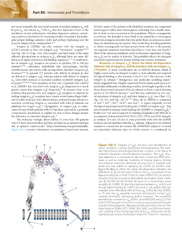
6 Figure 120–3. Diagram of α β structure and identification of
2 3 select mutations causing Glanzmann thrombasthenia. The web-
IIb 3
site http://med.mssm.edu/glanzmanndb contains a full listing of
2
reported Glanzmann thrombasthenia mutations. The α β struc-
IIb 3
7 ture depicted is a composite of data from crystal and NMR struc-
tures, as well as molecular modeling of missing regions. Among
the missense mutations identified are ones that (1) interfere with
inside-out and outside-in signaling (β S752P); (2) interfere with
3
ligand binding to either the metal ion-dependent adhesion site
5 (MIDAS) in β (β D119Y and D119N) or the α component of the
3
IIb
3
ligand binding site (Y143H, P145L/A, insert R160/T161); (3) result in
receptors that are sensitive to dissociation by divalent cation chela-
tion (β R214W, R214Q, R216Q); (4) result in a constitutively active
3
receptor (β C560R); (5) alter the interface between α and β and
3
3
IIb
4 disrupt ligand binding (β L262Y); (6) result in a β protein that can
3
3
complex more effectively with αV than α (S162L, R216Q, H280P);
IIb
or (7) alter the α propeller structure and prevent normal α β
IIb 3
IIb
complex formation, processing, and/or transport. The mutations
identified by number 8 in α (G991C and R995Q/W) and β (L718P
3
IIb
and D723H) are gain-of-function mutations associated with macro/
anisothrombocytopenia. (Reproduced with permission from Dr. Ana
Negri based on PDBids 3FCS, 3G9W, 2K9J, 2KNC, and 2KV9 and molec-
ular modeling of the missing segments of the α calf domain, the β3
IIb
hybrid domain, and the link between the β3 EGF-1 and EGF-2 domains.)
8 8
8
Kaushansky_chapter 120_p2039-2072.indd 2044 9/21/15 2:20 PM