
Page 2225 - Williams Hematology ( PDFDrive )
P. 2225
2200 Part XII: Hemostasis and Thrombosis Chapter 129: Disseminated Intravascular Coagulation 2201
PATHOGENESIS Endothelial perturbation constitutes a sine qua non for most
patients with DIC. Following injury or infection, the integrity of the
INFLAMMATION AND ENDOTHELIUM IN endothelium is compromised, mononuclear cells are activated by cytok-
DISSEMINATED INTRAVASCULAR ine and hormonal signals, additional cytokines and surface receptors
are upregulated, procoagulant proteins and platelets are activated, the
COAGULATION endothelium changes from an anticoagulant to procoagulant surface,
Various triggers cause an hemostatic imbalance that gives rise to a pro- and fibrinolysis is impeded. This sequence of events is typical for the
coagulant state (Fig. 129–1). The most important mediators responsi- systemic inflammatory response syndrome and can lead to microvascu-
ble for this imbalance are cytokines. There is an extensive crosstalk lar thrombosis with ensuing multiorgan dysfunction and eventually to
21
between coagulation and inflammatory systems, whereby inflammation multiorgan failure.
leads to activation of coagulation, and coagulation stimulates inflam-
matory activity. These interactions are highlighted in sepsis-induced ROLE OF CYTOKINES AND TISSUE FACTOR
22
systemic activation of coagulation and inflammation that lead to spe- Tissue factor (TF) plays a central role in the initiation of inflammation-
cific organ dysfunctions. The endothelium of the capillary bed is the induced coagulation in DIC. Blocking TF activity completely inhibits
23
28
most important interface in which the interaction between inflamma- inflammation-induced thrombin generation in experimental models of
tion and coagulation takes place. Endothelial cells may be a source of endotoxemia or bacteremia. 29,30 Most cells constitutively expressing TF
tissue factor and can thereby be involved in the initiation of coagulation are found in tissues not in direct contact with blood, such as the adven-
activation. All physiologic anticoagulant systems and various adhesion titial layer of larger blood vessels. TF becomes exposed to blood upon
molecules that may modulate both inflammation and coagulation are disruption of the vascular integrity, or when cells present in the circula-
connected to the endothelium. In sepsis, endothelial glycosaminogly- tion, such as monocytes, are triggered to express TF. The in vivo expres-
cans present in the glycocalyx are downregulated by proinflammatory sion of TF is dependent on interleukin (IL)-6 generation; inhibition of
cytokines, thereby impairing the functions of antithrombin (AT), tis- IL-6, unlike inhibition of other proinflammatory cytokines, completely
sue factor pathway inhibitor (TFPI), leukocyte adhesion, and leukocyte abrogates TF-dependent thrombin generation in experimental endotox-
transmigration. Because the glycocalyx also plays a role in other endo- emia. 21,31 In severe sepsis, monocytes, stimulated by proinflammatory
thelial functions, including maintenance of the vascular barrier func- cytokines, express TF, which leads to systemic activation of coagula-
tion, nitric oxide–mediated vasodilation, and antioxidant activity, all tion. Even in experimental low-dose endotoxemia in healthy subjects,
32
these processes can be impaired in DIC (see “Role of Oxidative Stress a 125-fold increase in TF mRNA levels in blood monocytes can be
and Vasoactive Molecules” below). 24,25 Moreover, specific disruption detected. A potential alternative source of TF may be endothelial cells,
33
of the glycocalyx results in thrombin generation and platelet adhesion polymorphonuclear cells, and other cell types. It is hypothesized that
within a few minutes. 26,27 TF from these sources is shuttled between cells through microparticles
derived from activated mononuclear cells. However, it is unlikely that
34
cells other than monocytes synthesize TF in substantial quantities. 32,35
Endothelial Tumor necrosis factor (TNF)-α and IL-1, also generated during inflam-
cells
mation, impair the physiologic anticoagulant pathways. 31,36,37
Cytokines Inflammatory cells
AMPLIFYING ROLE OF THROMBIN
AND PLATELETS
Tissue factor The TF–factor VIIa complex catalyzes the conversion of factor X to Xa,
expression
and factor Xa, in turn, forms the prothrombinase complex with fac-
tor Va, prothrombin (factor II), and calcium ions, thereby generating
Impairment of
physiologic thrombin, and converting fibrinogen into fibrin. The TF–factor VIIa
anticoagulant complex can also activate factor IX, and factor IXa forms the tenase
mechanisms complex with activated factor VIII and calcium ions, generating addi-
tional factor Xa, thereby forming an essential amplification loop of
Inhibition of thrombin generation. The assembly of the prothrombinase and tenase
fibrinolysis complexes are markedly facilitated if a suitable phospholipid surface is
because of available, such as the membrane of activated platelets. In the setting of
high inflammation-induced activation of coagulation, platelets can be acti-
levels of PAI-1
vated directly by endotoxin or by proinflammatory mediators, such as
the membrane of platelet-activating factor. Thrombin itself is one of the
Microvascular thrombosis & strongest platelet activators (Chap. 115).
modulation of inflammation Activation of platelets may also accelerate fibrin formation by
another mechanism. The expression of TF on monocytes is markedly
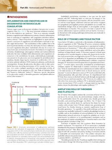
Figure 129–1. Schematic presentation of pathogenetic pathways stimulated by the presence of platelets and granulocytes in a P-selectin–
involved in the activation of coagulation in disseminated intravascular dependent reaction. This effect may be the result of nuclear factor
38
coagulation (DIC). In DIC, both perturbed endothelial cells and activated kappa B (NF-κB) activation induced by binding of activated platelets
mononuclear cells may produce proinflammatory cytokines that medi- to neutrophils and mononuclear cells. This cellular interaction also
39
ate coagulation activation. Activation of coagulation is initiated by tissue markedly enhances the production of IL-1β, IL-8, monocyte chemotac-
factor expression on activated mononuclear cells and endothelial cells. 40
In addition, downregulation of physiologic anticoagulant mechanisms tic protein (MCP)-1, and TNF-α.
and inhibition of fibrinolysis by endothelial cells further promote intra- Thrombin generated by the TF pathway amplifies both clotting and
vascular fibrin deposition. PAI-1, plasminogen-activator inhibitor type 1. inflammation through the following activities: (1) it activates platelets,
Kaushansky_chapter 129_p2199-2220.indd 2200 17/09/15 3:45 pm