
Page 2226 - Williams Hematology ( PDFDrive )
P. 2226
2200 Part XII: Hemostasis and Thrombosis Chapter 129: Disseminated Intravascular Coagulation 2201
giving rise to platelet aggregation and augmenting platelet functions activated by thrombin through cleavage of a specific aminoterminus bond
in coagulation; (2) it activates factors VIII, V, and XI, yielding fur- creating a tethered ligand that activates the receptor. PAR-2 can be cleaved
ther thrombin generation; (3) it activates proinflammatory factors via by factor Xa–TF–factor VIIa complex and by other proteases. Activated
48
protease-activated receptors (PARs); (4) it activates factor XIII to factor PARs then lead through mitogen-activated protein kinase and NF-κB
XIIIa, which crosslinks fibrin clots; (5) it activates thrombin-activatable signaling pathways to cell motility, shape change, proliferation, endoge-
fibrinolysis inhibitor (TAFI), making clots resistant to fibrinolysis; and nous secretagogue release, and apoptosis. The activated protein C (APC)–
(6) it increases expression of adhesion molecules, such as L-selectin, endothelial protein C receptor complex (see “Role of Natural Antico-
thereby promoting the inflammatory effects of leukocytes. 41 agulant Pathways” below) appears to be the “off switch” for PAR activa-
Paradoxically, at low concentrations, thrombin exhibits both anti- tion by the proteases. These counterbalances determine the magnitude
inflammatory and anticoagulant effects because it binds to thrombo- of coagulation and inflammatory upregulation by PARs. For exam-
modulin and activates protein C to the activated form, which, in turn, ple, factor VIIa–TF binding to PAR-2 in the lungs is proinflamma-
downregulates inflammation and serves as an “off switch” for further tory and appears to play a role in acute respiratory distress syndrome
thrombin generation (Chap. 116). (ARDS), raising the possibility that TFPI might be therapeutic in this
49
circumstance. This finding is consistent with data from animal stud-
ies demonstrating that TFPI can protect baboons from a LD100 of
ROLE OF COAGULATION PROTEASES IN Escherichia coli, likely by impeding factor VIIa–TF activation of PAR-2
UPREGULATING INFLAMMATION and thereby attenuating release of IL-6 and other proinflammatory agents.
Coagulation proteases and protease inhibitors not only interact with
coagulation proteins, but also with specific cell receptors to induce sig- ROLE OF FIBRINOGEN AND FIBRIN
naling pathways. In particular, protease interactions that affect inflam- Fibrinogen and fibrin directly influence the production of proinflamma-
matory processes may be important in critically ill patients. Coagulation tory cytokines and chemokines (including TNF-α, IL-1β, and MCP-1)
of whole blood in vitro results in a detectable expression of IL-1β mRNA by mononuclear cells and endothelial cells. Fibrinogen-deficient mice
50
in blood cells, and thrombin markedly enhances endotoxin-induced display inhibition of macrophage adhesion and less thrombin-mediated
42
IL-1 activity in culture supernatants of guinea pig macrophages. Simi- cytokine production in vivo. The effects of fibrinogen on mononuclear
43
larly, clotted blood produces IL-8 in vitro. 44 cells seem to be mediated by toll-like receptor-4, which is also the recep-
Factor Xa, thrombin, and fibrin can also activate endothelial cells, tor of endotoxin.
eliciting the synthesis of IL-6 and IL-8. 45,46 Coagulation proteases such
as thrombin, factor Xa, and factor VIIa–TF complex induce inflamma- ROLE OF NATURAL ANTICOAGULANT
tory upregulation via leukocyte, endothelial cell, and platelet PAR-1,
PAR-2, PAR-3, and PAR-4, which are located on leukocytes, endothelial PATHWAYS
47
cells, and platelets. PARs have an extracellular domain, seven trans- Procoagulant activity is regulated by three important anticoagulant
membrane domains, and an intracellular domain that is coupled to spe- pathways: AT, the protein C system, and TFPI. In DIC, the function of
cific G-proteins that transmit signaling. PAR-1, PAR-3, and PAR-4 are all three pathways can be impaired (Fig. 129–2). 51
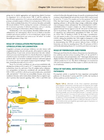
Figure 129–2. Schematic of the three important physiologic
anticoagulant mechanisms and their point of impact in the coagu-
Thrombomodulin
Protein C lation system. In sepsis, these mechanisms are impaired by various
mechanisms (green arrows). The protein C system is dysfunctional
as a result of low levels of zymogen protein C, downregulation of
Thrombin thrombomodulin and the endothelial protein C receptor, and low
levels of free protein S because of acute phase-induced high levels
of its binding protein (i.e., C4b-binding protein). There is a relative
insufficiency of the endothelial cell-associated tissue factor pathway
Activated Protein S inhibitor. The antithrombin system is defective because of low lev-
protein C Factor Va & els of antithrombin and impaired glycosaminoglycan expression on
factor VIIIa perturbed endothelial cells.
Endothelial
protein C receptor
Tissue factor
pathway inhibitor
Tissue factor
Glycosaminoglycans
Thrombin &
Antithrombin factor Xa
Kaushansky_chapter 129_p2199-2220.indd 2201 17/09/15 3:45 pm