
Page 2317 - Williams Hematology ( PDFDrive )
P. 2317
2290 Part XII: Hemostasis and Thrombosis Chapter 134: Atherothrombosis: Disease Initiation, Progression, and Treatment 2291
TABLE 134–2. Criteria for Defining the Vulnerable Plaque, TABLE 134–4. Pathophysiologic Differences Between
Based on the Study of Culprit Plaques Arterial and Venous Thrombi
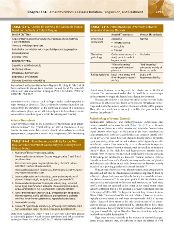
MAJOR CRITERIA Arterial Thrombosis Venous Thrombosis
Active Inflammation (monocyte/macrophage and sometimes Underlying Abnormal Normal
T-cell infiltration) vasculature • Atherosclerosis
Thin cap with large lipid core • Vasculitis
Endothelial denudation with superficial platelet aggregation • Trauma
Fissured plaque Thrombus Occlusive or nonocclu- Occlusive
Stenosis >90% pathology sive (mural thrombi in
MINOR CRITERIA large arteries)
Superficial calcified nodule “White thrombus” “Red thrombus”
Glistening yellow composed mainly of composed mainly of
platelets
fibrin, red cells
Intraplaque hemorrhage Pathophysiology Local shear stress and Stasis and
Endothelial dysfunction thrombogenic vascular hypercoagulability
Outward (positive) remodeling surface
Reproduced with permission from Naghavi M, Libby P, Falk E, et al:
From vulnerable plaque to vulnerable patient: A call for new defi-
nitions and risk assessment strategies: Part I. Circulation 2003 Oct clinical complications, including acute MI, stroke, and critical limb
7;108(14):1664-1672. ischemia. The previous section described in detail the current concepts
of the consecutive stages of atherosclerotic lesion development.
However, thrombosis is not simply the final occlusive event. It also
nonatherosclerotic disease, such as hypertrophic cardiomyopathy or contributes to atherosclerosis lesion development. Intraplaque hemor-
right ventricular dysplasia. Thus, a vulnerable patient should be con- rhage and in situ thrombosis localizes thrombin activity within plaques.
sidered from the standpoint of the combined presence of a vulnerable Thus, atheroma evolution is not only a proliferative process but also
atherosclerotic plaque, vulnerable blood (prone to thrombosis), and/or involves thrombosis. 162
vulnerable myocardium (prone to life-threatening arrhythmia).
Pathobiology of Arterial Thrombi
Arterial Thrombosis Fundamental pathologic and pathophysiologic distinctions exist
Atherothrombosis refers to the occurrence of thrombosis upon athero- between arterial and venous thrombi (Table 134–4). Arterial thrombi
sclerotic lesions, the typical setting for arterial thrombosis. It rep- usually are occlusive in smaller arteries and arterioles. Nonocclusive
161
resents the acute event that converts chronic atherosclerosis—a silent, mural thrombi often occur in the lumina of the heart chambers and
asymptomatic, progressive disease—into symptomatic, life-threatening
large arteries, such as the aorta and the iliac and common carotid arter-
ies. In any arterial vessel, however, thrombi develop almost invariably
TABLE 134–3. Blood Hypercoagulability Factors That upon preexisting abnormal intimal surfaces, which typically are ath-
May Contribute to Patient Vulnerability to Coronary Heart erosclerotic lesions. Less commonly, arterial thrombosis is superim-
Disease Events posed on other forms of vascular disease, such as vasculitis or traumatic
injury. Thus, in the high-flow and high-pressure arterial system,
163
1. Markers of blood hypercoagulability thrombi form in response to increased local shear forces and exposure
Decreased anticoagulation factors (e.g., proteins C and S and of thrombogenic substances on damaged vascular surfaces. Arterial
antithrombin) thrombi, referred to as white thrombi, are composed mainly of platelets
Prothrombotic gene polymorphisms (e.g., factor V Leiden, and relatively little fibrin or red cells. Leukocytes are likewise actively
G20210A prothrombin mutation) recruited into growing, platelet-rich arterial thrombi. 164
Increased coagulation factors (e.g., fibrinogen, factor VII, factor Thus, at sites of atherosclerotic plaque rupture, circulating platelets
VIII, von Willebrand factor) are activated not only by thrombogenic substances exposed to them by
2. Increased platelet activation (e.g., gene polymorphisms of a disrupted plaque but also directly by the locally increased shear forces
85
platelet integrin αI β , integrin α β , glycoprotein Ib/IX) the platelets encounter. At any given point in the circulation, shear
Ib 3 2 1
3. Decreased endogenous fibrinolysis activity (e.g., reduced rates are maximal adjacent to the vessel wall (measured as “wall shear
tissue-type plasminogen activator, increased plasminogen- rates”), and they are minimal in the center of the vessel lumen where
activator inhibitor (PAI)-1, certain PAI-1 polymorphisms) velocity of flowing blood is the greatest. Normally, wall shear rates are
–1
4. Other thrombogenic factors (e.g., anticardiolipin antibodies, in the range of 300 to 800 s in large arteries, and they increase to about
–1
thrombocytosis, sickle cell disease, polycythemia, diabetes 500 to 1600 s in arterioles of the microcirculation. However, in patho-
mellitus, hyperhomocysteinemia, hypercholesterolemia) logically stenotic vessels the wall shear rates can reach 10,000 s or even
–1
5. Increased viscosity higher. Increased shear stress in the microenvironment of an athero-
6. Transient hypercoagulability (e.g., smoking, dehydration, infec- sclerotic plaque is usually compounded by turbulent blood flow. These
tion, adrenergic surge, cocaine, estrogens, postprandial) locally abnormal hemodynamic forces can directly activate platelets as
they pass through the region. Disturbed flow can simultaneously cause
Data from Naghavi M, Libby P, Falk E, et al: From vulnerable plaque localized endothelial dysfunction. 165
to vulnerable patient: A call for new definitions and risk assessment High shear stresses, especially in the presence of marked shear gra-
strategies: Part I. Circulation 2003 Oct 7;108(14):1664-1672. dients around stenotic sites, are sufficient to cause the release of VWF
Kaushansky_chapter 134_p2281-2302.indd 2291 17/09/15 3:49 pm