
Page 570 - Williams Hematology ( PDFDrive )
P. 570
544 Part VI: The Erythrocyte Chapter 36: Pure Red Cell Aplasia 545
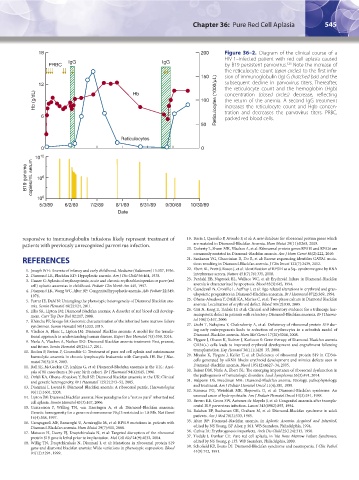
18 200 Figure 36–2. Diagram of the clinical course of a
HIV-1–infected patient with red cell aplasia caused
IgG IgG 123
PRBC by B19 persistent parvovirus. Note the increase of
the reticulocyte count (open circles) to the first infu-
150 sion of immunoglobulin (Ig) G (hatched bar) and the
12 µ subsequent decline in parvovirus titers. Thereafter,
the reticulocyte count and the hemoglobin (Hgb)
Hb (g/dL) Hb 100 Reticulocytes (1000/ L) concentration (closed circles) decrease, reflecting
the return of the anemia. A second IgG treatment
increases the reticulocyte count and Hgb concen-
6 tration and decreases the parvovirus titers. PRBC,
packed red blood cells.
50
Reticulocytes
0 0
12
B19 genome copies/mL serum 10 6
10
10 0
5/3/89 6/2/89 7/2/89 8/1/89 8/31/89 9/30/89 10/30/89
Date
responsive to immunoglobulin infusions likely represent treatment of 19. Boria I, Quarello P, Avondo F, et al: A new database for ribosomal protein genes which
patients with previously unrecognized parvovirus infection. are mutated in Diamond-Blackfan Anemia. Hum Mutat 29(11):E263, 2008.
20. Doherty L, Sheen MR, Vlachos A, et al: Ribosomal protein genes RPS10 and RPS26 are
commonly mutated in Diamond-Blackfan anemia. Am J Hum Genet 86(2):222, 2010.
REFERENCES 21. Sankaran VG, Ghazvinian R, Do R, et al: Exome sequencing identifies GATA1 muta-
tions resulting in Diamond-Blackfan anemia. J Clin Invest 122(7):2439, 2012.
1. Joseph WH: Anemia of infancy and early childhood. Medicine (Baltimore) 15:307, 1936. 22. Ebert BL, Pretz J, Bosco J, et al: Identification of RPS14 as a 5q− syndrome gene by RNA
2. Diamond LK, Blackfan KD: Hypoplastic anemia. Am J Dis Child 56:464, 1938. interference screen. Nature 451(7176):335, 2008.
3. Gasser C: Aplasia of erythropoiesis; acute and chronic erythroblastopenias or pure (red 23. Perdahl EB, Naprstek BL, Wallace WC, et al: Erythroid failure in Diamond-Blackfan
cell) aplastic anaemias in childhood. Pediatr Clin North Am 445, 1957. anemia is characterized by apoptosis. Blood 83(3):645, 1994.
4. Diamond LK, Wang WC, Alter BP: Congenital hypoplastic anemia. Adv Pediatr 22:349, 24. Casadevall N, Croisille L, Auffray I, et al: Age-related alterations in erythroid and gran-
1976. ulopoietic progenitors in Diamond-Blackfan anaemia. Br J Haematol 87(2):369, 1994.
5. Farrar JE, Dahl N: Untangling the phenotypic heterogeneity of Diamond Blackfan ane- 25. Ohene-Abuakwa Y, Orfali KA, Marius C, et al: Two-phase culture in Diamond Blackfan
mia. Semin Hematol 48(2):124, 2011. anemia: Localization of erythroid defect. Blood 105(2):838, 2005.
6. Ellis SR, Lipton JM: Diamond Blackfan anemia: A disorder of red blood cell develop- 26. Giri N, Kang E, Tisdale JF, et al: Clinical and laboratory evidence for a trilineage hae-
ment. Curr Top Dev Biol 82:217, 2008. matopoietic defect in patients with refractory Diamond-Blackfan anaemia. Br J Haema-
7. Khincha PP, Savage SA: Genomic characterization of the inherited bone marrow failure tol 108(1):167, 2000.
syndromes. Semin Hematol 50(4):333, 2013. 27. Uechi T, Nakajima Y, Chakraborty A, et al: Deficiency of ribosomal protein S19 dur-
8. Vlachos A, Blanc L, Lipton JM: Diamond Blackfan anemia: A model for the transla- ing early embryogenesis leads to reduction of erythrocytes in a zebrafish model of
tional approach to understanding human disease. Expert Rev Hematol 7(3):359, 2014. Diamond-Blackfan anemia. Hum Mol Genet 17(20):3204, 2008.
9. Narla A, Vlachos A, Nathan DG: Diamond Blackfan anemia treatment: Past, present, 28. Flygare J, Olsson K, Richter J, Karlsson S: Gene therapy of Diamond Blackfan anemia
and future. Semin Hematol 48(2):117, 2011. CD34(+) cells leads to improved erythroid development and engraftment following
10. Rodon P, Breton P, Courouble G: Treatment of pure red cell aplasia and autoimmune transplantation. Exp Hematol 36(11):1428–35, 2008.
haemolytic anaemia in chronic lymphocytic leukaemia with Campath-1H. Eur J Hae- 29. Miyake K, Flygare J, Keifer T, et al: Deficiency of ribosomal protein S19 in CD34+
matol 70(5):319, 2003. cells generated by siRNA blocks erythroid development and mimics defects seen in
11. Ball SE, McGuckin CP, Jenkins G, et al: Diamond-Blackfan anaemia in the U.K.: Anal- Diamond-Blackfan anemia. Blood 105(12):4627–34, 2005.
ysis of 80 cases from a 20-year birth cohort. Br J Haematol 94(4):645, 1996. 30. Raiser DM, Narla A, Ebert BL: The emerging importance of ribosomal dysfunction in
12. Orfali KA, Ohene-Abuakwa Y, Ball SE: Diamond Blackfan anaemia in the UK: Clinical the pathogenesis of hematologic disorders. Leuk Lymphoma 55(3):491, 2014.
and genetic heterogeneity. Br J Haematol 125(2):243–52, 2005. 31. Halperin DS, Freedman MH: Diamond-Blackfan anemia: Etiology, pathophysiology,
13. Dianzani I, Loreni F: Diamond-Blackfan anemia: A ribosomal puzzle. Haematologica and treatment. Am J Pediatr Hematol Oncol 11(4):380, 1989.
93(11):1601, 2008. 32. Scimeca PG, Weinblatt ME, Slepowitz G, et al: Diamond-Blackfan syndrome: An
14. Lipton JM: Diamond blackfan anemia: New paradigms for a “not so pure” inherited red unusual cause of hydrops fetalis. Am J Pediatr Hematol Oncol 10(3):241, 1988.
cell aplasia. Semin Hematol 43(3):167, 2006. 33. Brown KE, Green SW, Antunez de Mayolo J, et al: Congenital anaemia after transpla-
15. Gustavsson P, Willing TN, van Haeringen A, et al: Diamond-Blackfan anaemia: cental B19 parvovirus infection. Lancet 343(8902):895, 1994.
Genetic homogeneity for a gene on chromosome 19q13 restricted to 1.8 Mb. Nat Genet 34. Balaban EP, Buchanan GR, Graham M, et al: Diamond-Blackfan syndrome in adult
16(4):368, 1997. patients. Am J Med 78(3):533, 1985.
16. Campagnoli MF, Ramenghi U, Armiraglio M, et al: RPS19 mutations in patients with 35. Alter BP: Diamond-Blackfan anemia, in Aplastic Anemia, Acquired and Inherited,
Diamond-Blackfan anemia. Hum Mutat 29(7):911, 2008. edited by NS Young, BP Alter, p 361. WB Saunders, Philadelphia, 1994.
17. Matsson H, Davey EJ, Draptchinskaia N, et al: Targeted disruption of the ribosomal 36. Cathie IA: Erythrogenesis imperfecta. Arch Dis Child 25(124):313, 1950.
protein S19 gene is lethal prior to implantation. Mol Cell Biol 24(9):4032, 2004. 37. Tisdale J, Dunbar CE: Pure red cell aplasia, in The Bone Marrow Failure Syndromes,
18. Willig TN, Draptchinskaia N, Dianzani I, et al: Mutations in ribosomal protein S19 edited by NS Young, p 135. WB Saunders, Philadelphia, 2000.
gene and diamond blackfan anemia: Wide variations in phenotypic expression. Blood 38. Schofield KP, Evans DI: Diamond-Blackfan syndrome and neutropenia. J Clin Pathol
94(12):4294, 1999. 44(9):742, 1991.
Kaushansky_chapter 36_p0539-0548.indd 545 9/17/15 6:15 PM