
Page 578 - Williams Hematology ( PDFDrive )
P. 578
552 Part VI: The Erythrocyte Chapter 37: Anemia of Chronic Disease 553
TABLE 37–2. Laboratory Studies of Iron Metabolism in Iron-Deficiency Anemia and Anemia of Inflammation
IDA (n = 48) AI (n = 58) COMBI (n = 17)
Hemoglobin, g/L 93 ± 16 (96) 102 ± 12 (103) 88 ± 20 (90)
MCV, fL 75 ± 9 (75) 90 ± 7 (91) 78 ± 9 (79)
Iron, μmol/L (10–40) 8 ± 11 (4) 10 ± 6 (9) 6 ± 3 (6)
Transferrin, g/L (2.1–3.4 m, 3.3 ± 0.4 (3.3) 1.9 ± 0.5 (1.8) 2.6 ± 0.6 (2.4)
2.0–3.1 f)
Transferrin saturation, % 12 ± 17 (5.7) 23 ± 13 (21) 12 ± 7 (8)
Ferritin, μg/L (15–306 m, 5–103 f) 21 ± 55 (11) 342 ± 385 (195) 87 ± 167 (23)
TfR, mg/L (0.85–3.05) 6.2 ± 3.5 (5.0) 1.8 ± 0.6 (1.8) 5.1 ± 2.0 (4.7)
TfR/log ferritin 6.8 ± 6.5 (5.4) 0.8 ± 0.3 (0.8) 3.8 ± 1.9 (3.2)
f, Females; m, males; TfR, transferrin receptor.
Diagnosis was defined by marrow iron stain and appropriate coexisting disease. Patients with a combination of no stainable marrow iron and
either coexisting disease or elevated CRP were classified as “COMBI.” Normal ranges for this laboratory for males (m) and females (f) are indi-
cated. Measurements are presented as mean ± SD (median).
Modified with permission from Punnonen K, Irjala K, Rajamaki A: Serum transferrin receptor and its ratio to serum ferritin in the diagnosis of iron
deficiency. Blood 89(3):1052–1057, 1997.
number of sideroblasts is characteristic of AI. Although marrow stain DIFFERENTIAL DIAGNOSIS
could be considered the gold standard for differential diagnosis of AI
and iron deficiency, the discomfort to the patient associated this proce- Most patients with chronic infections, inflammatory diseases, or neo-
dure, reports of variability in interpretation and the wide availability plastic disorders are anemic. The diagnosis of AI should only be made if
70
of the serum ferritin assay have decreased the use of marrow stain in the anemia is mild to moderate, the serum iron and iron-binding capac-
this setting. ity are low, and the serum ferritin is elevated. Anemia of CKD is rare in
mild renal disease but common and often severe in end-stage renal dis-
ease. Underlying diseases, comorbidities, and their treatments can cause
many types of anemia, so other potential causes should be considered.
Serum ferritin
1. Drug-induced marrow suppression or drug-induced hemolysis can
2500 2500 complicate infections, inflammatory disorders, CKDs, and cancer.
When the marrow is suppressed by cytotoxic drugs or idiopathic
1000 1000 toxic reaction, serum iron tends to be high and reticulocyte count
low. In hemolysis, reticulocyte counts, haptoglobin, bilirubin, and
lactate dehydrogenase often are elevated.
2. Chronic blood loss depletes iron stores and decreases serum iron
300 300
and serum ferritin but increases transferrin (Chap. 43). When AI
and chronic blood loss coexist, serum ferritin usually indicates the
100 100 predominant disorder, although the level can increase as a result
of inflammation itself. Chronic blood loss from hemodialysis or
mcg/L 50 50 mcg/L occult gastrointestinal bleeding is common in anemia of CKD and
its lowering effect on ferritin may be masked by coexisting inflam-
mation. Testing stool for occult blood and looking for other sources
of overlooked blood loss, including phlebotomy, urinary loss and
15 15
menorrhagia, often identify the source of bleeding. Once this issue is
addressed, a successful trial of iron repletion confirms the diagnosis
5 5 of iron deficiency complicating AI or anemia of CKD.
3. Endocrine disorders, including hypothyroidism and hyperthyroid-
ism, testicular failure, and diabetes mellitus, can be associated with
a chronic normocytic, normochromic anemia (Chap. 38). Unless
inflammation or associated iron deficiency is present, serum iron
1 1 should be normal in these disorders.
IDA COMBI ACD 4. Anemia resulting from metastatic invasion of the marrow by tumors
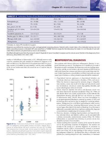
Figure 37–3. Distribution of serum ferritin measurements in patients can be the presenting symptom of malignancy. The anemia can
with iron-deficiency anemia (IDA), anemia of chronic disease (ACD = develop in the setting of a previous diagnosis of carcinoma or
anemia of inflammation [AI]) and combined IDA and ACD (COMBI).
The horizontal lines indicate lower normal values for healthy men and lymphoma and by itself is accompanied by normal or increased
women. (Reproduced with permission from Punnonen K, Irjala K, Rajamaki serum iron (Chap. 45). It often develops in the setting of preexist-
A: Serum Transferrin Receptor and Its Ratio to Serum Ferritin in the Diagnosis ing malignancy-related AI. The blood film often is abnormal, with
of Iron Deficiency. Blood 89(3):1052–1057, 1997.) poikilocytes, teardrop-shaped red cells, normoblasts, or immature
Kaushansky_chapter 37_p0549-0558.indd 553 9/17/15 6:17 PM