
Page 665 - Williams Hematology ( PDFDrive )
P. 665
640 Part VI: The Erythrocyte Chapter 43: Iron Deficiency and Overload 641
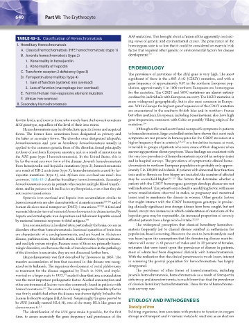
TABLE 43–3. Classification of Hemochromatosis HFE mutations. This brought about a fusion of the apparently contrast-
ing views of genetic and environmental causes. The penetrance of the
I. Hereditary Hemochromatosis homozygous state is so low that it could be considered an essential risk
A. Classical hemochromatosis (HFE hemochromatosis) (type 1) factor that required other genetic or environmental factors for disease
B. Juvenile hemochromatosis (type 2) development. 213
1. Abnormality in hemojuvelin
2. Abnormality of hepcidin EPIDEMIOLOGY
C. Transferrin receptor-2 deficiency (type 3) The prevalence of mutations of the HFE gene is very high. The most
D. Ferroportin abnormalities (type 4) significant of these is the c.845 A→G (C282Y) mutation, and with a
1. Gain of function (systemic iron overload) gene frequency of approximately 0.07 in the northern European pop-
2. Loss of function (macrophage iron overload) ulation, approximately 5 in 1000 northern Europeans are homozygous
E. Ferritin H-chain iron-responsive element mutation for the mutation. The C282Y and S65C mutations are almost entirely
F. African iron overload confined to individuals with European ancestry. The H63D mutation is
more widespread geographically, but is also most common in Europe-
II. Secondary Hemochromatosis
ans. Within Europe the highest gene frequencies of the C282Y mutation
are encountered in the southern British Isles and in northern France
but other northern Europeans, including Scandinavians, also have high
ferritin levels, and even to those who merely have the hemochromatosis gene frequencies, consistent with Celtic or possibly Viking origin of the
HFE genotype, regardless of the level of their iron stores. mutation. 214
Hemochromatosis may be divided into genetic forms and acquired Although earlier studies attributed nonspecific symptoms in patients
forms. The former have sometimes been designated as primary and to hemochromatosis, large controlled series have shown that most such
the latter as secondary forms. The disorder once designated idiopathic symptoms are not present in homozygotes for the C282Y mutation at a
hemochromatosis and now as hereditary hemochromatosis usually is higher frequency than in controls, 215–217 or a borderline increase, at most,
applied to the common genetic form of the disorder, found principally invariably in groups of patients who were aware of their diagnosis when
in those of northern European ancestry, and as a result of mutations in answering questions about symptoms. These findings are consistent with
the HFE gene (type I hemochromatosis). In the United States, this is the very low prevalence of hemochromatosis reported in autopsy series
by far the most common form of the disease. Juvenile hemochromatosis and in hospital surveys. The prevalence of symptomatic clinical hemo-
from hemojuvelin and hepcidin mutations (type 2), hemochromatosis chromatosis in northern European populations is probably only approx-
as a result of TfR-2 mutations (type 3), hemochromatosis caused by fer- imately 5 in 100,000 individuals. If patients with abnormal liver function
roportin mutations (type 4), and African iron overload are much less tests and/or fibrosis on liver biopsy are included, the number of affected
common. Table 43–3 classifies hereditary hemochromatosis. Secondary may be severalfold higher. 218–221 The factors that determine whether a
hemochromatosis occurs in patients who receive multiple blood transfu- patient with the C282Y homozygous genotype develops disease are not
sions, and in patients with ineffective erythropoiesis, even when they do well understood. The patient’s sex is clearly a modifying factor, with more
not receive transfusions. severe manifestations observed in males, as pregnancy and menstrual
Systemic iron overload and hepatic iron accumulation similar to losses tend to ameliorate the disease in women. Other genetic factors
hemochromatosis are also characteristic of atransferrinemia 203–206 and of that might interact with the C282Y homozygous genotype in produc-
human divalent metal transporter (DMT)-1 mutations. 207,208 A fetal and ing clinically significant iron storage disease have been sought, but not
neonatal disorder termed neonatal hemochromatosis is characterized by found, except rare instances in which coinheritance of mutations of the
hepatic and extrahepatic iron deposition and fulminant hepatitis caused hepcidin gene may be responsible. An increased proportion of severely
by maternal immune response to fetal antigens. 209 affected patients have a large alcohol intake. 222,223
Iron accumulation in localized sites, particularly the brain, occurs in The widespread perception that classical hereditary hemochro-
disorders other than hemochromatosis. Increased quantities of brain iron matosis frequently led to clinical disease resulted in enthusiasm for
are characteristic of a ceruloplasminemia, and are found in Alzheimer population-based screening. However, the cost-to-benefit analysis used
disease, parkinsonism, Friedreich ataxia, Hallervorden-Spatz syndrome, was based upon the assumptions that life-threatening disease manifes-
and multiple system atrophy. Because none of these are primarily hema- tations will occur in 43 percent of males and in 28 percent of females,
tologic disorders, and because the role of iron deposition in the pathology estimates that were based upon the prevalence of disease in patients,
of the disorders is uncertain, they are not discussed further here. most of whom had been diagnosed clinically with hemochromatosis.
Hemochromatosis was first described by Trousseau in 1865. The With the realization that the clinical penetrance is much lower, interest
massive accumulation of iron that occurred in this disease was recog- in screening the general population for hemochromatosis has largely
nized as its hallmark. The ingenious development of serial phlebotomy disappeared.
as treatment for the disease suggested by Finch in 1949, and imple- The prevalence of other forms of hemochromatosis, including
mented on a larger scale in 1952, made it clear that iron accumulation juvenile hemochromatosis, hemochromatosis as a result of ferroportin
210
was the most important pathogenetic factor. Alcohol consumption and deficiency, and atransferrinemia, is much lower than that the prevalence
other environmental factors were also commonly found in patients with of classical hereditary hemochromatosis. These forms of hemochroma-
hemochromatosis. The existence of a long-suspected hereditary factor tosis are very rare.
211
was firmly established when the disease was shown tightly linked to the
human leukocyte antigen (HLA locus). Surprisingly, the gene proved to
be HFE (initially named HLA-H), one of the many HLA-like genes on ETIOLOGY AND PATHOGENESIS
chromosome 6. 212 Toxicity of Iron
The identification of the HFE gene made it possible, for the first In living organisms, iron associates with proteins to function in oxygen
time, to assess accurately the gene frequency and penetrance of the storage and transport and in various metabolic reactions as an electron
Kaushansky_chapter 43_p0627-0650.indd 640 9/17/15 6:27 PM