
Page 661 - Williams Hematology ( PDFDrive )
P. 661
636 Part VI: The Erythrocyte Chapter 43: Iron Deficiency and Overload 637
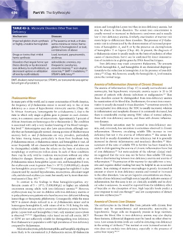
TABLE 43–2. Microcytic Disorders Other Than Iron minor, and hemoglobin Lepore trait than in iron-deficiency anemia, but
may be absent in these disorders. The serum iron concentration is
Deficiency
usually normal or increased in thalassemic syndromes and is usually
Mechanisms Diseases low in iron-deficiency anemia. Similarly, examination of marrow iron
Impaired globin chain synthesis β-Thalassemia or trait, α-thalas- stores helps to differentiate these disorders. The presence of β-thalas-
or highly unstable hemoglobin semia minima or minor, hemo- semia trait is substantiated by the demonstration of increased propor-
globin H, hemoglobin E or trait, tions of hemoglobin A and F, or by the presence on electrophoresis
2
combinations of above of hemoglobin H or Lepore (Chap. 48). At present, the diagnosis of
Drugs or toxins that inhibit Lead, isoniazid, pyrazinamide, α-thalassemia minor is usually made on the basis of exclusion of other
heme synthesis sirolimus causes of microcytosis, but it can be confirmed by direct demonstra-
tion of mutations in α-globin genes by DNA-based techniques.
Disorders that impair heme syn- sideroblastic anemias, ery- Iron deficiency may mask concurrent thalassemia. The amounts
thesis directly or by decreased thropoietic porphyrias,
iron delivery to erythroblasts, or atransferrinemia, aceruloplas- of both hemoglobin A and hemoglobin H are diminished dispropor-
203
2
decreased uptake or utilization minemia, DMT-1 mutations, tionately to the reduction in hemoglobin A in the presence of iron defi-
207
284
182
of iron by erythroblasts STEAP3 deficiency 285 ciency (Chap. 46); however, usually the hemoglobin A level remains
2
above the normal range.
DMT, divalent metal transporter; STEAP3, six-transmembrane epithe-
lial antigen of prostate 3.
Anemia of Inflammation (Anemia of Chronic Disease)
The anemia of inflammation (Chap. 37) is usually normochromic and
normocytic, but hypochromic microcytic anemia occurs in 20 to 30
139
percent of patients with chronic infections or malignancies. Thus
Thalassemia Minor these disorders cannot be distinguished from iron-deficiency anemia
In many parts of the world, and in many communities of North America, by examination of the blood film. Furthermore, the serum iron concen-
139
the frequency of β-thalassemia minor is second only to that of iron tration is usually decreased in these disorders, sometimes severely. In
deficiency as a cause of hypochromic microcytic anemia (Chap. 48). uncomplicated iron deficiency, the TIBC is usually increased, whereas
In African Americans, homozygosity for α-thalassemia-2, that is the in inflammatory and neoplastic diseases it is commonly decreased, but
state in which only single α-globin gene is present on each chromo- there is considerable overlap among TIBC values of normal subjects,
some, is a common cause of microcytosis. Approximately 3 percent of those with iron-deficiency anemia, and those with chronic inflamma-
African Americans are homozygous for α-thalassemia-2. The condition tory diseases.
is associated with only a very modest lowering of the blood hemoglo- Transferrin saturation may be normal in iron-deficiency ane-
bin level. Heterozygotes may also have microcytosis, although usu- mia, and, conversely, low saturation is sometimes observed in chronic
177
ally they are hematologically normal. Among persons of Mediterranean inflammation. However, circulating soluble TfRs increase in iron
ancestry both α- and β-thalassemia are very prevalent, particularly deficiency but not in the anemia of inflammation. The serum fer-
170
the latter. Among Asians, particularly in those from Southeast Asia, ritin level is usually diminished in iron deficiency, but it is generally
β-thalassemia minor, α-thalassemia minor, and hemoglobin E trait, all increased in chronic inflammatory and neoplastic disorders. Mea-
147
occur frequently. All are characterized by microcytosis, and none can surement of the ratio of soluble TfR to ferritin has been found to be
be distinguished reliably from the others on the basis of erythrocyte useful in distinguishing the anemia of chronic inflammation from that
170
morphology or erythrocyte indices alone. In each of these conditions of iron deficiency, but meta-analysis of the relevant clinical stud-
there may be only mild to moderate microcytosis without any other ies suggested that the ratio may not be better than soluble TfR assay
distinctive changes. However, in the majority of patients with α- or alone at discriminating between iron deficiency anemia and anemia of
183
β-thalassemia minor, hemoglobin Lepore trait, and hemoglobin E trait, inflammation. Examination of the marrow for stainable iron is inva-
the erythrocyte count is greater than 5 × 10 /L (5,000,000/μL), despite sive and requires skilled reading but may be helpful in an occasional
12
low hemoglobin concentration. 178,179 Homozygous hemoglobin E is also patient. Iron staining of marrow macrophages is greatly decreased in
characterized by marked hypochromia, microcytosis, abundant target amount or absent in iron deficiency anemia and normal or increased
cells, and elevated erythrocyte count, but usually not by more than min- in the other disorders. Low serum hepcidin concentrations are charac-
imal anemia (Chap. 49). 178 teristic of iron deficiency and high serum hepcidin indicates anemia of
In contrast to the findings in these hemoglobinopathies, ery- inflammation, but this assay is not yet clinically available and its clini-
throcyte counts of 5 × 10 /L (5,000,000/μL) or higher are relatively cal value is unknown. As would be expected from the inhibitory effect
12
uncommon among adults with iron-deficiency anemia. However, of hepcidin on the absorption of iron, high hepcidin levels predict a
180
184
erythrocytosis may be seen in children with iron-deficiency anemia or poor response to oral iron therapy and low incorporation of dietary
in polycythemia vera patients who have become iron deficient following iron into erythrocytes. 185
hemorrhage or therapeutic phlebotomy. Consequently, while the mean
MCV is almost always reduced in α- or β-thalassemia minor and in Anemia of Chronic Liver Disease
homozygous hemoglobin E, with values of 60 to 70 fL being the rule, The erythrocytes in the blood film from patients with chronic liver
values this low are seen only in severe iron-deficiency anemia. In hemo- disease may be normochromic and normocytic, macrocytic, or
globin Lepore trait and hemoglobin E trait, only minimal microcytosis hypochromic. Target cells are frequently present in large numbers.
is observed. 178,179,181 Algorithmic rules based on red cell counts, MCV Because the blood film in iron-deficiency anemia may also display
or RDW are not sufficiently reliable for distinguishing iron deficiency these features, differential diagnosis must be based on other observa-
from thalassemia in populations with high prevalence of iron deficiency tions. Low serum ferritin levels are useful in detecting iron deficiency
compared to thalassemias. in the setting of cirrhosis, but normal or even increased serum fer-
186
Mild reticulocytosis, polychromatophilia, and basophilic stippling are ritin does not exclude iron deficiency, especially in the presence of
more likely to be encountered in β-thalassemia minor, δβ-thalassemia active liver injury. 186,187
Kaushansky_chapter 43_p0627-0650.indd 636 9/17/15 6:27 PM