
Page 683 - Williams Hematology ( PDFDrive )
P. 683
658 Part VI: The Erythrocyte Chapter 45: Anemia Associated with Marrow Infiltration 659
TABLE 45–1. Causes of Marrow Infiltration contribute to anemia: elevated hepcidin (Chap. 37) and other factors,
including hematopoiesis-inhibiting cytokines released from tumor cells
I. Fibroblasts and collagen (Chap. 37) and iron (Chap. 42) and folate and cobalamin (Chap. 41)
A. Primary myelofibrosis (PMF) deficiencies. When they are excluded, the finding discussed above sug-
gests that only massive marrow replacement leads to anemia.
B. Fibrosis associated with other myeloproliferative neo-
plasms (MPNs)
C. Fibrosis of hairy cell leukemia CLINICAL FEATURES
D. Metastatic tumors (e.g., breast carcinoma) Symptoms and signs associated with infiltrative marrow disorders usu-
E. Sarcoidosis 14,15 ally are related to the underlying disease. Other symptoms, such as
fatigue, often from upregulated cytokines, may also contribute to ane-
F. Secondary myelofibrosis with pulmonary hypertension
mia itself. Some patients are asymptomatic, and the incidental discovery
II. Other noncellular material of cytopenias and leukoerythroblastic blood morphology leads to diag-
A. Oxalosis 6 nosis of an underlying disorder
III. Tumor cells
A. Carcinomas (breast, lung, prostate, kidney, LABORATORY FEATURES
thyroid and neuroblastoma) 7,8,11 BLOOD
B. Sarcoma 10
The anemia usually is mild to moderate, but it can be severe. White
IV. Granulomas 14 cell and platelet counts may vary, but the most characteristic feature is
A. Sarcoidosis the morphologic appearance of red cells on the blood film. These cells
may show anisocytosis and poikilocytosis, but the presence of teardrop
B. Fungal infections
forms and nucleated red cells is particularly suggestive of marrow infil-
C. Miliary tuberculosis tration (Chap. 31; Fig. 45–2). The combination of nucleated red cells
V. Macrophages and immature myeloid precursors constitutes the leukoerythroblastic
picture that is characteristic of marrow infiltration and extramedullary
A. Gaucher disease
hematopoiesis. The presence of cancer cells on the blood film occurs
B. Niemann-Pick disease 16 occasionally and always indicates marrow invasion (Fig. 45–3). 27
C. Macrophage activation syndrome (MAS) 34,35
VI. Marrow necrosis MARROW
A. Sickle cell anemia 19 Marrow biopsy is the most reliable procedure used to diagnose
marrow-infiltrative disease and should be performed in all patients
B. Solid tumor metastasis 18
with suspected metastatic carcinoma or hematologic features of mye-
C. Septicemia 18 lophthisic anemia. Marrow aspiration 24,28 does not provide a reliable
D. Acute lymphoblastic leukemia yield of tumor cells and is particularly difficult in primary or second-
ary myelofibrosis. The inability to aspirate marrow (dry tap) leads to
E. Arsenic therapy 22
a high degree of suspicion of marrow replacement and accompany-
VII. Failure of osteoclast development ing myelofibrosis. Because the diagnostic marrow yield from biopsies
A. Osteopetrosis 36 depends on the amount of tissue examined, bilateral posterior iliac crest
marrow biopsies may be necessary. In patients with metastatic cancer
A B
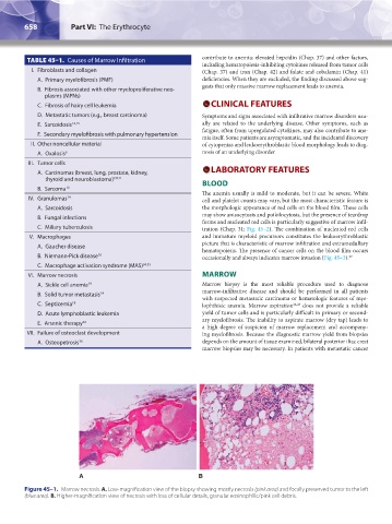
Figure 45–1. Marrow necrosis. A. Low-magnification view of the biopsy showing mostly necrosis (pink area) and focally preserved tumor to the left
(blue area). B. Higher-magnification view of necrosis with loss of cellular details, granular eosinophilic/pink cell debris.
Kaushansky_chapter 45_p0657-0660.indd 658 9/17/15 6:40 PM