
Page 687 - Williams Hematology ( PDFDrive )
P. 687
662 Part VI: The Erythrocyte Chapter 46: Erythrocyte Membrane Disorders 663
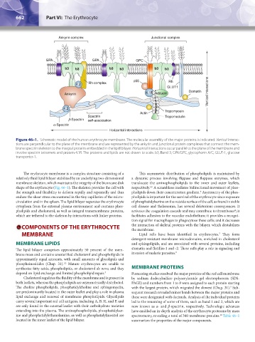
Ankyrin complex Junctional complex
GPA GPA GPC
b3 b3 b3 b3 b3 b3
p55 Rh
4.2 Rh complex
GLUT1
Ankyrin 4.1R Adducin Dematin Vertical interactions
4.2
Actin Tropomyosin
Spectrin Tropomodulin
b-Spectrin self-association
a-Spectrin
Horizontal interactions
Figure 46–1. Schematic model of the human erythrocyte membrane. The molecular assembly of the major proteins is indicated. Vertical interac-
tions are perpendicular to the plane of the membrane and are represented by the ankyrin and junctional protein complexes that connect the mem-
brane spectrin skeleton to the integral proteins embedded in the lipid bilayer. Horizontal interactions occur parallel to the plane of the membrane and
involve spectrin tetramers and protein 4.1R. The proteins and lipids are not drawn to scale. b3, Band 3; GPA/GPC, glycophorin A/C; GLUT-1, glucose
transporter-1.
The erythrocyte membrane is a complex structure consisting of a This asymmetric distribution of phospholipids is maintained by
relatively fluid lipid bilayer stabilized by an underlying two-dimensional a dynamic process involving flippase and floppase enzymes, which
membrane skeleton, which maintains the integrity of the biconcave disk translocate the aminophospholipids to the inner and outer leaflets,
shape of the erythrocyte (Fig. 46–1). The skeleton provides the cell with respectively. A scramblase mediates bidirectional movement of phos-
4,5
the strength and flexibility to deform rapidly and repeatedly and thus pholipids down their concentration gradient. Asymmetry of the phos-
6
endure the shear stress encountered in the tiny capillaries of the micro- pholipids is important for the survival of the erythrocyte since exposure
circulation and in the spleen. The lipid bilayer separates the erythrocyte of phosphatidylserine on the outside surface of the cell, as found in sickle
cytoplasm from the external plasma environment and contains phos- cell disease and thalassemia, has several deleterious consequences. It
pholipids and cholesterol, as well as integral transmembrane proteins, activates the coagulation cascade and may contribute to thromboses ; it
4
which are tethered to the skeleton by interactions with linker proteins. facilitates adhesion to the vascular endothelium; it provides a recogni-
tion signal for macrophages to phagocytose these cells; and it decreases
COMPONENTS OF THE ERYTHROCYTE the interaction of skeletal proteins with the bilayer, which destabilizes
the membrane.
MEMBRANE Lipid rafts have been identified in erythrocytes. They form
7
detergent-resistant membrane microdomains, enriched in cholesterol
MEMBRANE LIPIDS and sphingolipids, and are associated with several proteins, including
The lipid bilayer comprises approximately 50 percent of the mem- stomatin and flotillin-1 and -2. These rafts play a role in signaling and
brane mass and contains unesterified cholesterol and phospholipids in invasion of malaria parasites. 8
approximately equal amounts, with small amounts of glycolipids and
phosphoinositides (Chap. 31). Mature erythrocytes are unable to
1,2
synthesize fatty acids, phospholipids, or cholesterol de novo, and they MEMBRANE PROTEINS
depend on lipid exchange and limited phospholipid repair. 3 Pioneering studies resolved the major proteins of the red cell membrane
Cholesterol regulates the fluidity of the membrane and is present in by sodium dodecylsulfate polyacrylamide gel electrophoresis (SDS-
both leaflets, whereas the phospholipids are asymmetrically distributed. PAGE) and numbers from 1 to 8 were assigned to each protein starting
The choline phospholipids, phosphatidylcholine and sphingomyelin, with the largest protein, which migrated the slowest (Chap. 31). Sub-
9
are predominantly located in the outer leaflet and play a role in plasma sequent research revealed minor bands between the major proteins and
lipid exchange and renewal of membrane phospholipids. Glycolipids these were designated with decimals. Analysis of the individual proteins
carry several important red cell antigens, including A, B, H, and P, and led to the renaming of some of them, such as band 1 and 2, which are
are only found in the external leaflet with their carbohydrate moieties now known as α- and β-spectrin, respectively. Technologic advances
extending into the plasma. The aminophospholipids, phosphatidylser- have enabled an in-depth analysis of the erythrocyte proteome by mass
ine and phosphatidylethanolamine, as well as phosphatidylinositol are spectrometry, revealing a total of 340 membrane proteins. Table 46–1
10
located in the inner leaflet of the lipid bilayer. summarizes the properties of the major components.
Kaushansky_chapter 46_p0661-0688.indd 662 9/17/15 6:41 PM