
Page 684 - Williams Hematology ( PDFDrive )
P. 684
658 Part VI: The Erythrocyte Chapter 45: Anemia Associated with Marrow Infiltration 659
A B
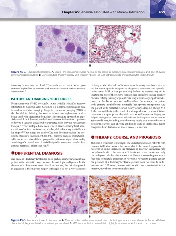
Figure 45–2. Leukoerythroblastosis. A. Blood film containing several nucleated red blood cells (RBCs), few circulating blasts, and RBCs showing
severe anisopoikilocytosis. B. Corresponding marrow biopsy with reticulin fibrosis (3+) and intrasinusoidal megakaryocytes (black arrows).
involving the marrow, the blood CD34-positive cell count can be up to technique, with the help of immunocytochemistry and flow cytome-
50 times higher than in patients with metastatic cancer without marrow try for tumor-specific antigens, its diagnostic sensitivity and specific-
involvement. 29 ity increases. MRI or isotopic scanning before the marrow may aid in
locating the site of the biopsy. Hematologic disorders causing marrow
ISOTOPE AND IMAGING PROCEDURES fibrosis, notably primary myelofibrosis, may mimic a myelophthisic dis-
order, but the distinctions are usually evident. For example, the patient
99m
Technetium-99m ( Tc) sestamibi uptake reliably identifies marrow with primary myelofibrosis invariably has splenic enlargement and
infiltration by Gaucher cells. Sestamibi is a pharmaceutical agent used the patient with metastatic cancer nearly always does not (Chap. 91).
in nuclear medicine imaging. Magnetic resonance imaging (MRI) is If the myelophthisis is the result of a storage disease or other infiltra-
also helpful for defining the severity of marrow replacement and is tive cause, the appropriate chemical tests, as well as marrow biopsy, are
being used with increasing frequency. This imaging approach is espe- helpful in diagnosis. Nucleated red cells and leukocytosis can be seen in
cially useful for following resolution of marrow infiltration in patients acute conditions, including overwhelming sepsis, acute severe hypoxia,
with type 1 Gaucher disease who are treated with enzyme-replacement postcardiac arrest, and chronic conditions such as thalassemia major,
therapy. 20,30,31 An isotopic bone scan or MRI study showing focal accu- congestive heart failure, and severe hemolytic anemia.
mulation of radioactive tracers can be helpful in locating a suitable site
for biopsy, 20,32 but a negative study of the area does not exclude the pos-
sibility of marrow involvement. On MRI, marrow necrosis characteristi- THERAPY, COURSE, AND PROGNOSIS
cally has an extensive, diffuse, geographic pattern of signal abnormality
consisting of a central area of variable signal intensity surrounded by a The goal of treatment is managing the underlying disease. Patients with
distinct peripheral enhancing rim. 21 marrow infiltration caused by cancer should be treated appropriately;
however, in some instances the presence of marrow infiltration may
DIFFERENTIAL DIAGNOSIS not adversely affect the outcome. If treatment is successful, not only
the malignant cells but also the reactive fibrosis surrounding metastatic
The cause of a leukoerythroblastic blood picture is known to occur in a foci may completely disappear. In hormone-refractory prostate cancer,
patient with metastatic cancer or overt hematologic malignancy. In the the presence of a leukoerythroblastic picture does not seem to influ-
33
absence of a likely cause after clinical evaluation, the initial approach ence survival. However, in most patients with cancers metastatic to the
to diagnosis is the marrow biopsy. Although it is not a very sensitive marrow, only short-term survival is a rule.
A B
Figure 45–3. Metastatic tumor in the marrow. A. Marrow packed with melanoma cells and displacing normal marrow elements. Tumor cells have
characteristic large nuclei with prominent pink nucleoli. B. S100 immunohistochemical stain highlights melanoma infiltrates in the marrow.
Kaushansky_chapter 45_p0657-0660.indd 659 9/17/15 6:40 PM