
Page 873 - Williams Hematology ( PDFDrive )
P. 873
848 Part VI: The Erythrocyte Chapter 55: Alloimmune Hemolytic Disease of the Fetus and Newborn 849
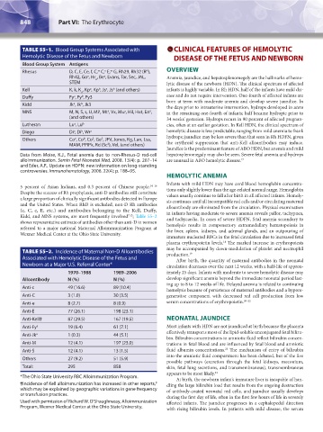
TABLE 55–1. Blood Group Systems Associated with CLINICAL FEATURES OF HEMOLYTIC
Hemolytic Disease of the Fetus and Newborn DISEASE OF THE FETUS AND NEWBORN
Blood Group System Antigens
Rhesus D, C, E, Ce, f, C, C, E, G, Rh29, Rh32 (R ), OVERVIEW
w
N
x
w
Rh42, Go , Hr , Be , Evans, Tar, Sec, JAL, Anemia, jaundice, and hepatosplenomegaly are the hallmarks of hemo-
a
a
o
STEM lytic disease of the newborn (HDN). The clinical spectrum of affected
Kell K, k, K , Kp , Kp , Js , Js (and others) infants is highly variable. In Rh HDN, half of the infants have mild dis-
b
a
b
a
o
Duffy Fy , Fy , Fy3 ease and do not require intervention. One-fourth of affected infants are
b
a
born at term with moderate anemia and develop severe jaundice. In
Kidd Jk , Jk , Jk3 the days prior to intrauterine intervention, hydrops developed in utero
b
a
MNS M, N, S, s, U, Mi , Mt , Vx, Mur, Hil, Hut, En , in the remaining one-fourth of infants; half became hydropic prior to
a
a
a
(and others) 34 weeks’ gestation. Hydrops recurs in 90 percent of affected pregnan-
Lutheran Lu , Lu b cies, often at an earlier gestation. In Kell HDN, the clinical spectrum of
a
Diego Di , Di , Wr a hemolytic disease is less predictable, ranging from mild anemia to frank
b
a
hydrops; jaundice may be less severe than that seen in Rh HDFN, given
a
Others Co , Co , Co , Ge , JFV, Jones, Kg, Lan, Lsa, the erythroid suppression that anti-Kell alloantibodies may induce.
b
3
3
MAM, PPIPk, Rd (Sc ), Vel, (and others)
4
Jaundice is the predominant feature of ABO HDN, but anemia and mild
Data from Moise, K.J., Fetal anemia due to non-Rhesus-D red-cell hepatosplenomegaly may also be seen. Severe fetal anemia and hydrops
alloimmunization. Semin Fetal Neonatal Med, 2008. 13(4): p. 207–14 are unusual in ABO hemolytic disease. 17
and Eder, A.F., Update on HDFN: new information on long-standing
controversies. Immunohematology, 2006. 22(4): p. 188–95.
HEMOLYTIC ANEMIA
Infants with mild HDN may have cord blood hemoglobin concentra-
5 percent of Asian Indians, and 0.3 percent of Chinese people. 14–16 tions only slightly lower than the age-related normal range. Hemoglobin
Despite the success of Rh prophylaxis, anti-D antibodies still constitute values usually continue to fall after birth in all affected infants. Hemoly-
a large proportion of clinically significant antibodies detected in Europe sis continues until all incompatible red cells and/or circulating maternal
and the United States. When RhD is excluded, non-D Rh antibodies alloantibody are eliminated from the circulation. Physical examination
(c, C, e, E, etc.) and antibodies belonging to the Kell, Duffy, in infants having moderate to severe anemia reveals pallor, tachypnea,
7,13
Kidd, and MNS systems, are most frequently involved ; Table 55–2 and tachycardia. In cases of severe HDFN, fetal anemia secondary to
shows representative estimate of antibodies other than anti-D in women hemolysis results in compensatory extramedullary hematopoiesis in
referred to a major national Maternal Alloimmunization Program at the liver, spleen, kidneys, and adrenal glands, and an outpouring of
Wexner Medical Center at the Ohio State University.
immature nucleated RBCs in the fetal circulation due to increased fetal
plasma erythropoietin levels. The marked increase in erythropoiesis
18
may be accompanied by down-modulation of platelet and neutrophil
TABLE 55–2. Incidence of Maternal Non-D Alloantibodies production. 19
Associated with Hemolytic Disease of the Fetus and After birth, the quantity of maternal antibodies in the neonatal
Newborn at a Major U.S. Referral Center* circulation decreases over the next 12 weeks, with a half-life of approx-
1970–1988 1989–2006 imately 25 days. Infants with moderate to severe hemolytic disease may
Alloantibody N (%) N (%) develop significant anemia beyond the immediate neonatal period last-
ing up to 8 to 12 weeks of life. Delayed anemia is related to continuing
Anti-c 49 (16.6) 89 (10.4)
hemolysis because of persistence of maternal antibodies and a hypore-
Anti-C 3 (1.0) 30 (3.5) generative component with decreased red cell production from low
Anti-e 8 (2.7) 8 (0.9) serum concentrations of erythropoietin. 20–22
Anti-E 77 (26.1) 198 (23.1)
Anti-Kell† 87 (29.5) 167 (19.5) NEONATAL JAUNDICE
Anti-Fy a 19 (6.4) 61 (7.1) Most infants with HDN are not jaundiced at birth because the placenta
effectively transports most of the lipid-soluble unconjugated fetal biliru-
Anti-Jk a 1 (0.3) 44 (5.1)
bin. Bilirubin concentrations in amniotic fluid reflect bilirubin concen-
Anti-M 12 (4.1) 197 (23.0) trations in fetal blood and are influenced by fetal blood and amniotic
23
Anti-S 12 (4.1) 13 (1.5) fluid albumin concentrations. The mechanism of entry of bilirubin
into the amniotic fluid compartment has been debated, but of the five
Others 27 (9.2) 51 (5.9)
possible pathways (excretion through the fetal kidneys, meconium,
Total: 295 858 skin, fetal lung secretions, and transmembranous), transmembranous
appears to be most likely. 24
*The Ohio State University RBC Alloimmunization Program. At birth, the newborn infant’s immature liver is incapable of han-
8
†Incidence of Kell alloimmunization has increased in other reports, dling the large bilirubin load that results from the ongoing destruction
which may be explained by geographic variations in gene frequency of antibody-coated neonatal red cells, and jaundice usually develops
or transfusion practices. during the first day of life, often in the first few hours of life in severely
Used with permission of Richard W. O’Shaughnessy, Alloimmunization affected infants. The jaundice progresses in a cephalopedal direction
Program, Wexner Medical Center at the Ohio State University. with rising bilirubin levels. In patients with mild disease, the serum
Kaushansky_chapter 55_p0847-0862.indd 848 9/18/15 11:52 PM