
Page 877 - Williams Hematology ( PDFDrive )
P. 877
852 Part VI: The Erythrocyte Chapter 55: Alloimmune Hemolytic Disease of the Fetus and Newborn 853
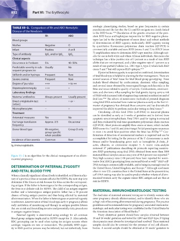
TABLE 55–3. Comparison of Rh and ABO Hemolytic serologic phenotyping studies, based on gene frequencies in certain
populations and the fact that the C/c and E/e antigens are closely linked
Disease of the Newborn
to the RHD locus. 14,56 Elucidation of the genetic structure of the prev-
Rh ABO alent RHD locus and haplotypes responsible for RhD-negative pheno-
Blood groups types has led to the development of more direct and robust methods
of determination of RHD zygosity molecularly. RHD zygosity testing
Mother Negative O by quantitative fluorescence polymerase chain reaction (QF-PCR) is
Infant Positive A or B commercially available and uses RHD (exons 5 and 7) to RHCE (exon
7) amplification ratios to determine RHD copy number. Although suit-
Type of antibody IgG and/or IgG IgG
1 3 2 able for use in both white and ethnic African individuals, this molecular
Clinical aspects technique has a false-positive rate of 1 percent as a result of rare RHD
Occurrence in firstborn 5% 40–50% alleles that are not expressed, and a false-negative rate of 1 percent as a
result of rare partial D alleles (i.e., DBT type 1, type 2) which lack RHD
Predictable severity in sub- Usually No 69,70
sequent pregnancies exons 5 and 7 but still express RhD epitopes.
If paternal heterozygosity is suspected or confirmed, determination
Stillbirth and/or hydrops Frequent Rare of fetal blood type is helpful in planning further management. There are
Severe anemia Frequent Rare several sources of fetal tissue for fetal blood group genotyping. These
Degree of jaundice +++ + to ++ include blood obtained by cordocentesis, chorionic villus sampling,
and cervical tissue obtained by transvaginal lavage; each has risks to the
Hepatosplenomegaly +++ + fetus and issues related to quality of sample. Cordocentesis, amniocen-
Laboratory findings tesis, and chorionic villus sampling for fetal genetic typing carry a risk
Maternal antibodies Always present Usually present of FMH with increased risk of augmenting maternal sensitization and of
fetal loss. 45,46 The advent of noninvasive methods of prenatal diagnosis
Direct antiglobulin test + + or − using fetal DNA extracted from maternal plasma as early as the first tri-
(infant) mester of pregnancy has obviated those concerns and has dramatically
Microspherocytes − + improved the ability to perform molecular testing on fetal tissue. 71
Treatment Circulating cell-free fetal DNA (ccff-DNA) in maternal plasma
can be identified as early as 5 weeks of gestation and is derived from
Antenatal measures Yes No apoptotic syncytiotrophoblasts. Fetal DNA used for typing is extracted
Exchange transfusion Approx. 2/3 Occasional and then evaluated by real-time quantitative polymerase chain reaction
frequency (QT-PCR). Most protocols amplify three exons or more, which include
Donor blood type Rh-negative, Group O only RHD exons 4 to 7 and 10, and detect target Psi (ψ) pseudogene sequences
group specific in exon 4 to avoid false-positives when the fetus has RHDψ. 72,73 Con-
when possible firmation of detection of nonmaternal markers is required and can be
accomplished by testing for the presence of the Y chromosome in male
Incidence of late anemia Common Rare
fetuses and/or housekeeping genes such as hemoglobin β-chain, β-
Ig, immunoglobulin. actin, albumin, or chemokine receptor 5. A recent meta-analysis
reviewed 37 publications describing 44 protocols reporting noninva-
sive RHD genotyping using fetal DNA obtained from more than 3000
72
Figure 55–2 is an algorithm for the clinical management of an alloim- maternal blood samples; an accuracy rate of 94.8 percent was reported.
munized pregnancy. Very high accuracy rates (>96 percent) have been reported for nonin-
74
vasive fetal RHCE genotyping from maternal blood as well. RHD ccff-
DETERMINATION OF PATERNAL ZYGOSITY DNA testing is commercially available, and is being increasingly used in
75
the United States, United Kingdom, and Europe. Although used more
AND FETAL BLOOD TYPE often in non-U.S. countries than in the United States at the present time,
When a clinically significant alloantibody is identified, or if there is a his- ccff-DNA testing may also be used to predict whether infants of alloim-
tory of a previous fetus or neonate affected by HDFN, the next step is to munized women carry the cognate minor RBC antigen and are thus at
determine if the fetus is at risk because the fetus carries the correspond- risk for HDFN.
ing antigen. If the father is homozygous for the corresponding antigen,
the fetus is at definite risk for HDFN. The child of an antigen-negative
mother and a heterozygous antigen-positive father has a 50 percent MATERNAL IMMUNOHEMATOLOGIC TESTING
chance of being antigen-positive and thus being affected by maternal The dual aims of maternal antenatal testing are to identify women who
alloimmunization. When the father is heterozygous or paternal zygosity enter pregnancy already alloimmunized, and to identify those who are
is unknown, determination of fetal blood type early in pregnancy allows at high risk of becoming alloimmunized during pregnancy. The practice
early institution of monitoring and therapy in antigen-positive fetuses guidelines and recommendations for pregnancy-associated immunohe-
that are at risk and forestalling invasive and potentially risky procedures matologic and molecular testing were established in the United States by
in antigen negative fetuses. the American Association of Blood Banks. 57
Paternal zygosity is determined using serology for all common Every obstetrical patient should have samples obtained between
blood group antigens implicated in HDFN except for D. Alternatively, 10 and 16 weeks’ gestation and tested for ABO and RhD type; D typing
RBC genotyping can be used when typing for antigen systems where discrepancies must always be investigated and resolved. These maternal
serologic reagents are rare or nonexistent. The probable RHD zygos- samples should also be screened for the presence of red cell alloanti-
ity in RhD-positive persons may be inferred, but not definitively, from bodies. A second sample should be obtained at 28 weeks’ gestation to
Kaushansky_chapter 55_p0847-0862.indd 852 9/18/15 11:52 PM