
Page 876 - Williams Hematology ( PDFDrive )
P. 876
850 Part VI: The Erythrocyte Chapter 55: Alloimmune Hemolytic Disease of the Fetus and Newborn 851
of IgG and IgG subclasses, the relative contribution of each of these Kell HDFN. 59–61 In contrast to the hemolysis observed in RhD HDFN,
3
1
subclasses to the severity of HDFN remains controversial. 51,54,55 the severe fetal anemia observed because of maternal anti-Kell alloan-
tibodies is largely a result of suppression of erythropoiesis and not to
HEMOLYTIC DISEASE CAUSED BY OTHER RED hemolysis. Clinical observations of inappropriately low levels of cir-
CELL ANTIBODIES culating reticulocytes and normoblasts for the degree of anemia have
Although antibodies against RhD tend to be the most clinically sig- long been noted in affected fetuses, and suppression of erythropoiesis
has been established by in vitro studies showing that growth of Kell-
nificant in terms of fetal outcomes, there are many other RBC antigens positive erythroid progenitor cells is inhibited by monoclonal IgG and
capable of inducing alloantibodies after exposure through transfu- IgM anti-Kell antibodies. Furthermore, anti-Kell antibodies are asso-
62
sion or pregnancy. Any alloantibody capable of inducing hemolysis or ciated with suppression of megakaryocyte and granulocyte colony-
suppressing erythropoiesis may be clinically significant to developing forming units with resultant fetal and neonatal thrombocytopenia and
fetuses. However, the mere presence of antibodies on screening tests pancytopenia. 63,64 As discussed in further detail later in this the section”
may not be clinically significant, because of the unique characteristics MATERNAL IMMUNOHEMATOLOGIC TESTING”, titers of maternal
b
a
of some antibodies. For example, antibodies to Lewis antigens (Le , Le ) anti-Kell do not necessarily correlate with the severity of fetal anemia
are IgM and do not cross the placenta. Alternatively, Lutheran (Lu , Lu ) and thus all maternal anti-Kell alloantibodies must be considered to be
a
b
and Chido antigens are poorly expressed on fetal and neonatal red cells potentially clinically significant to antigen positive fetuses.
and therefore are not susceptible to destruction by maternal antibodies.
Case reports may be biased toward more-severe cases, and there
is considerable variability in the clinical spectrum of disease produced Other Minor Antigens (Non-D, Non-Kell)
by different alloantibodies. With the widespread use of RhIg, anti-Kell Many other minor RBC antigens besides D and Kell are immunogenic
has bypassed anti-D as the leading cause of HDFN in some centers. in transfusion and pregnancy settings, with some of these alloantibodies
8
Table 55–1 lists some of the antibodies more commonly associated with having the capacity of being detrimental to developing fetuses. The inci-
HDFN. 13,56 Of note, many cases severe enough to require IUT involve dence and prevalence of other minor antigens contributing to HDFN
antibodies to RhD, with or without antibodies to other alloantigens depends in part on the geographical area evaluated, as genetics and local
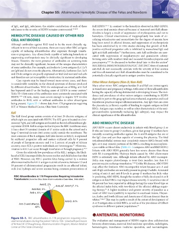
being present. Figure 55–1 shows data from 178 pregnancies requiring transfusion practices impact alloimmunization. Any IgG that can cross
IUT at Wexner Medical Center, Ohio State University. the placenta is, in theory, capable of binding to cognate antigen on fetal
RBCs. Antigen copy number, as well as other characteristics of the anti-
Kell gens/antibodies (potentially including IgG subtype), may impact the
The Kell blood group system consists of at least 28 discrete antigens, of clinical significance of the alloantibodies.
which eight are associated with HDFN. The KEL gene is located on chro-
mosome 7q34, and Kell antigens are located on the red cell membrane gly- ABO HEMOLYTIC DISEASE
coprotein CD238. Kell is unique in that it spans the RBC membrane once; ABO HDN occurs almost exclusively in infants with blood group A or
it has a short N-terminal domain of 47 amino acids in the cytosol and a B who are born to group O mothers, given that group O mothers have
large C-terminal domain (665 amino acids) outside the membrane. The naturally occurring antibodies against the A and B antigens that are of
most common of the K antigens, Kell (also known as KEL1), is expressed the IgG class and are thus capable of crossing the placenta. The fetal
by erythroid progenitor cells and mature erythroid cells by 9 percent reticuloendothelial system may completely remove RBCs bound with
of people of European ancestry and 1 to 2 percent of people of African IgG, or it may remove portions of the RBCs, resulting in microsphero-
ancestry; most KEL1-positive individuals are heterozygous. Alloimmu- cytes visible on blood film. (Table 55–3 compares ABO and RhD HDN.)
57
nization to KEL1 can occur through transfusion or through pregnancy. 58,59 Infants with ABO HDN generally have less-severe disease than those
Given the relatively low prevalence of the KEL1 antigen, the likeli- with Rh incompatibility. Hydrops fetalis caused by ABO alloimmune
hood of KEL1 incompatibility between mother and child is less than that HDN is extremely rare. Although infants affected by ABO incompat-
of RhD. However, any KEL1-positive fetus being carried by a woman ibility may require phototherapy to treat their jaundice, less than 0.1
alloimmunized to the KEL1 antigen is at risk of anemia. Between 2.5 and percent require exchange transfusion. 17,65 Unlike Rh disease, ABO HDN
10 percent of alloimmunized pregnancies result in an affected infant, may affect the firstborn ABO-incompatible infant because anti-A and
with fetal hydrops and severe anemia being common presentations of anti-B IgG antibodies are normally present in group O adults. Antenatal
testing of anti-A and anti-B levels in group O mothers has little value
RBC Alloantibodies in 178 Pregnancies Requiring Intrauterine in predicting ABO HDN, though the number of fully developed A or B
Transfusions (from the Ohio State Alloimmunization Committee, 1965-2007) antigens on fetal RBCs may impact disease severity. A recurrence rate of
88 percent has been reported in siblings having the same blood type as
Anti-E, 2%
a
b
Anti-Js , Anti-Fy , 1% the affected index baby, with two-thirds of the affected siblings requir-
Anti-Kell, 5% ing therapy. A higher incidence and greater severity of jaundice as a
34
Anti-c, 6% result of ABO incompatibility is reported in southeast Asians, Hispan-
ics, Arabs, and South African and Americans of African descent than in
whites. 17,66,67 This may be partly a result of the extent of development of
Anti-D, 44% A or B antigen sites on fetal RBCs, as well as of the prevalence of Gilbert
Anti-D and other(s), 68
42% syndrome in different patient populations.
ANTENATAL MONITORING
Figure 55–1. RBC alloantibodies in 178 pregnancies requiring intra-
uterine transfusions during the period 1965 to 2007. (Used with permission The evaluation and management of HDFN require close collaboration
of Richard W. O’Shaughnessy, Alloimmunization Program, Wexner Medical between obstetricians, maternal fetal medicine specialists, radiologists,
Center at Ohio State University.) hematologists, transfusion medicine specialists, and neonatologists.
Kaushansky_chapter 55_p0847-0862.indd 851 9/18/15 11:52 PM