
Page 891 - Williams Hematology ( PDFDrive )
P. 891
866 Part VI: The Erythrocyte Chapter 56: Hypersplenism and Hyposplenism 867
TABLE 56–2. Causes of Massive Splenomegaly patients develop marked blood cytopenias is not clear, although folate
deficiency is a factor in some instances. The presence of thrombocy-
1. Myeloproliferative disorders topenia or leukopenia in patients with chronic liver disease is associated
a. Primary myelofibrosis with increased mortality. 37
b. Chronic myeloid leukemia Ultrasound-guided fine-needle biopsy of the spleen can be use-
2. Lymphomas ful in circumstances in which the spleen holds the tissue required for
a. Hairy cell leukemia diagnosis, such as splenic lymphoma. However, fine-needle aspiration
is rarely a definitive diagnostic tool but can indicate monoclonality of
b. Chronic lymphocytic leukemia (especially prolymphocytic splenic lymphocytes, which is helpful and forces further diagnostic
variant) evaluation. Aspiration cytology and core biopsy can be obtained with
3. Infectious relative safety in experienced hands using image-guided fine needles. 38
a. Malaria The response to transfusion of blood products, especially platelets,
b. Leishmaniasis (kala azar) may be significantly impaired in patients with massive splenomegaly. 39
4. Extramedullary hematopoiesis
a. Thalassemia major THERAPY, COURSE, AND PROGNOSIS
5. Infiltrative Total Splenectomy
a. Gaucher disease Splenectomy is indicated as an emergency procedure after abdominal
trauma and partial rupture of the spleen. It also may be indicated when
splenic size or infarcts causes sustained left upper abdominal pain or dis-
The diagnosis of splenoptosis may be made coincidentally on an imaging comfort. Splenectomy has been used for the treatment of functionally
39
study. The condition may be accompanied by signs of hypersplenism, significant blood cytopenias. In such circumstances, case reports have
31
hyposplenism, and often, when developing slowly, is initially mistaken described dramatic restoration of blood counts to normal levels within
for a pelvic or lower abdominal tumor. days to weeks after splenectomy; however, the only controlled trial evalu-
ating relief of cytopenias showed no improvement. Orthotopic liver trans-
6
LABORATORY FEATURES plant corrects the cytopenias in the majority of patients with cirrhosis. 40
Hereditary spherocytosis, immune thrombocytopenic purpura,
The characteristic features of hypersplenism are splenomegaly, blood and immune hemolytic anemia are the most common indications for
cytopenias, and absence of other causes of cytopenias (e.g., anemia splenectomy. Splenectomy exerts its effect in autoimmune cytopenias
caused by bleeding). The blood cell morphology usually is normal, by improving cell survival and also by decreasing autoantibody pro-
although a few spherocytes may result from metabolic conditioning of duction. In thalassemia major, an improvement in the anemia is well
red cells during repeated slow transits through the expanded red pulp. described after splenectomy. In such cases, splenectomy may improve
Tests, such as epinephrine mobilization, were used in the past to try the response to transfusion. Some children with sickle cell anemia may
to distinguish sequestration from ineffective cellular production, but benefit from splenectomy if repeated sequestration crises with abdom-
results are difficult to interpret as epinephrine also releases platelets and inal pain occur before autosplenectomy renders the spleen atrophic. 41
neutrophils from marginal pools. 32 Splenectomy in patients with a massive spleen size (>1500 g), espe-
Thrombocytopenia is a common finding in patients with hepatic cially in primary myelofibrosis, is accompanied by higher morbidity and
cirrhosis, portal hypertension, and splenomegaly. In a retrospective mortality than is removal of the spleen for immune blood cytopenia.
42
study, 64 percent of patients with nonalcoholic cirrhosis had thrombo- Possible postoperative complications include extensive adhesions with
cytopenia. Other studies have found that approximately one-third of collateral blood vessels, hepatic or portal vein thrombosis, injury to the tail
33
patients with cirrhosis develop severe thrombocytopenia or neutrope- of the pancreas, operative site infections, and subdiaphragmatic abscesses.
nia. 34,35 Decompensated liver disease and history of alcohol consump- Laparoscopic splenectomy performed by experienced surgeons for
tion are independent risk factors for hypersplenism, but why some suitable hematologic conditions can result in less abdominal trauma
36
A B C
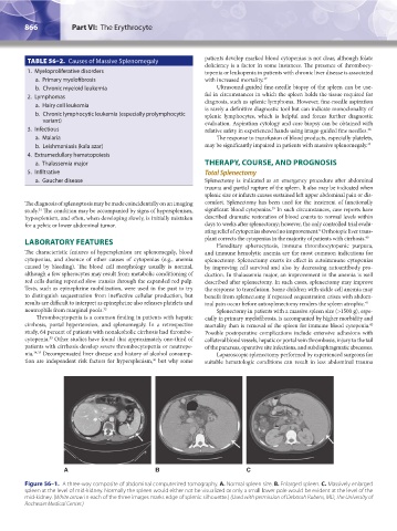
Figure 56–1. A three-way composite of abdominal computerized tomography. A. Normal spleen size. B. Enlarged spleen. C. Massively enlarged
spleen at the level of mid-kidney. Normally the spleen would either not be visualized or only a small lower pole would be evident at the level of the
mid-kidney. (White arrow in each of the three images marks edge of splenic silhouette.) (Used with permission of Deborah Rubens, MD, The University of
Rochester Medical Center.)
Kaushansky_chapter 56_p0863-0870.indd 866 9/17/15 3:05 PM