
Page 893 - Williams Hematology ( PDFDrive )
P. 893
868 Part VI: The Erythrocyte Chapter 56: Hypersplenism and Hyposplenism 869
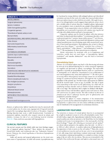
TABLE 56–3. Conditions Associated with Hyposplenism be functional in young children with enlarged spleens and disordered
circulation and may be the result of atrophy after repeated infarcts have
MISCELLANEOUS destroyed splenic tissue in older children and adults. Although the pres-
Surgical splenectomy ence of an enlarged spleen usually suggests hypersplenism, spleen size is
not a reliable index of splenic function. Complete splenic replacement
Splenic irradiation
by cysts, neoplastic tissue, or amyloid is an example of hyposplenic sple-
Sickle hemoglobinopathies nomegaly. Acute sequestration crises in children with sickle hemoglo-
64
Congenital agenesis binopathies, and occasionally in patients with malaria, may clog the red
cell pulp with cellular debris and lead to hyposplenism. 65,66
Thrombosis of splenic artery or vein
Congenital asplenia may be found in infants with situs inversus
Normal infants and other developmental abnormalities. Autoimmune disorders, such
38
67
GASTROINTESTINAL AND HEPATIC DISEASES as glomerulonephritis, systemic lupus erythematosus, 68,69 and rheuma-
70
toid arthritis, are occasionally associated with laboratory evidence and
Celiac disease
clinical manifestations (overwhelming sepsis with encapsulated bacte-
Dermatitis herpetiformis ria) of functional hyposplenism. Hyposplenism also occurs in chronic
73
Inflammatory bowel disease graft-versus-host disease, 71,72 sarcoidosis, alcoholic liver cirrhosis, 74,75
hepatic amyloidosis, 76,77 celiac disease, 78,79 and inflammatory bowel dis-
Cirrhosis
ease. 80,81 The mechanisms for these associations are unknown.
AUTOIMMUNE DISORDERS Splenic replacement by neoplastic cells, as in lymphomas and
Systemic lupus erythematosus leukemias, usually does not cause hyper- or hyposplenism. Splenic
82
irradiation and vascular obstruction may also lead to functional
83
Rheumatoid arthritis
hyposplenism.
Vasculitis
Glomerulonephritis Overwhelming Sepsis
Absence of a functional spleen may lead to life-threatening infections
Hashimoto thyroiditis
by removal of an efficient filtering bed in which opsonized organisms
Sarcoidosis are engulfed and destroyed by splenic macrophages. The responsible
HEMATOLOGIC AND NEOPLASTIC DISORDERS organism is typically an encapsulated bacteria, such as S. pneumoniae,
N. meningitidis, or H. influenzae. Unrestrained in vivo proliferation of
Graft versus host disease
such microorganisms may cause fatal septicemia. 84–86 The risk is great-
Essential thrombocytosis est among infants whose general immunologic system has not matured
Chronic lymphocytic leukemia enough to counteract bacterial infections, although the risk is present
regardless of the patient’s age. For this reason, splenectomy in children
Non-Hodgkin lymphoma
should be deferred until 5 years of age, if possible. The risk of sepsis
Hodgkin lymphoma varies depending on the reason for the splenectomy. In a child with an
Amyloidosis underlying immune disorder, such as Wiskott-Aldrich syndrome, the
risk is very high. The infectious risk is higher in children with thalas-
Advanced breast cancer
semia than in those with hereditary spherocytosis and lowest in those
Hemangiosarcoma with splenectomy for splenic trauma. The risk is reduced by the use of
SEPSIS/INFECTIOUS DISEASES pneumococcal and H. influenzae vaccines prior to splenectomy and
prophylactic penicillin therapy. 87
Malaria
Because the spleen is a major component of the mononuclear
Disseminated meningococcemia phagocyte system and has substantial lymphatic tissue in the white pulp,
hyposplenism or splenectomy can also reduce antibody synthesis that
may be beneficial in the management of autoimmune disorders.
disease), or splenectomy. Splenic hypofunction may be associated with
a normal spleen size. In some cases, engorgement of ingested materials
impairs the macrophage-dependent functions of the spleen. Impaired LABORATORY FEATURES
filtering function may cause a mild thrombocytosis. Functional or The reduction or absence of normal splenic function is accompanied
anatomical asplenia, especially after surgical removal in infants and by a slight to moderate increase in white cell and platelet counts. How-
children, increases the risk of an overwhelming bacterial infection. ell-Jolly bodies, target cells, Pappenheimer (siderotic) bodies, and occa-
Table 56–3 lists conditions associated with hyposplenism. sional acanthocytes often are present in the blood film, but the finding
of pitted erythrocytes in wet preparations is the most specific of all the
blood findings. Target cells reflecting an increased red cell surface
88
89
CLINICAL FEATURES are almost always present in the asplenic state, but only 1 in 100 to 1
The normal neonate and the elderly adult may have findings sugges- in 1000 red cells is affected. A sensitive indication of hyposplenism is
tive of impaired splenic function. These include the presence of the appearance of pits or pocks on the cell surface. These pits consist
90
60
Howell-Jolly bodies and erythrocyte pits (see “Laboratory Features” of submembranous vacuoles and can be seen only in wet preparations
below). However, the clinical significance of functional hyposplenism of red cells using direct interference-contrast microscopy. Intracellu-
is uncertain. 61–63 lar vesiculation containing hemoglobin is a normal occurrence during
Sickle cell anemia and surgical splenectomy are the most com- aging of the red cell in the circulation. This process is intensified in the
mon causes of hyposplenism. In sickle cell anemia, hyposplenism may last half of the erythrocyte life span and leads to a decreased mean cell
Kaushansky_chapter 56_p0863-0870.indd 868 9/17/15 3:05 PM