
Page 917 - Williams Hematology ( PDFDrive )
P. 917
892 Part VI: The Erythrocyte Chapter 58: The Porphyrias 893
the foundation for the development of animal models of this disorder
using halogenated polyaromatic hydrocarbons. 12,13 Strand and cowork-
ers described the enzyme deficiency in AIP for the first time in 1970, N N HO Cl N
14
and Bonkovsky and coworkers first reported treatment of a porphyria N N N N N N
15
patient with hemin in 1971. In the past several decades, the enzymes Fe 2+ Fe 3+ Fe 3+ Fe 2+
of the heme biosynthetic pathway have been defined in terms of their N N
amino acid composition, genomic and complementary DNA (cDNA) N N N N N N N
sequences, and crystal structures. Erythroid-specific and housekeeping
transcripts have been described for at least four enzymes in the pathway,
and progress made in understanding the regulation of heme synthesis
in specific tissues, especially the marrow and liver. Multiple mutations Pyridine
have been described in each of the human porphyrias, and some specific Hemin Hematin Hemin-HCl hemochrome
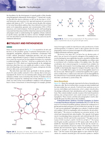
treatments introduced. Figure 58–3. Forms of iron protoporphyrin IX. The porphyrin macro-
cycle is represented only by its pyrrole nitrogen atoms.
ETIOLOGY AND PATHOGENESIS
HEME hemochromogen is useful for identification and quantification of heme
Heme (iron protoporphyrin IX; Fig. 58–2) is essential for all cells and and hemoproteins. In medicine, hemin is also a generic term for heme
preparations used as intravenous therapies for acute porphyrias, such as
functions as the prosthetic group of numerous hemoproteins such as lyophilized hematin and heme arginate.
hemoglobin, myoglobin, respiratory cytochromes, cytochromes P450 The ferrous iron atom (Fe ) in heme has six electron pairs, of
2+
(CYPs), catalase, peroxidase, tryptophan pyrrolase, and nitric oxide which four are bound to the pyrrolic nitrogens of the porphyrin mac-
synthase. Approximately 85 percent of heme is synthesized in the mar- rocycle, leaving two unoccupied electron pairs, one above and the other
row to meet the requirement for hemoglobin formation; the remainder below the plane of the porphyrin ring. In hemoglobin, one of these pairs
is synthesized largely in the liver. Most heme synthesized in the liver is coordinated with a histidine residue of the globin chain. The other
16
is required for CYPs, which are located primarily in the endoplasmic coordination site in deoxyhemoglobin is protected from oxidation by
reticulum where they turn over rapidly and oxidize a variety of chemi- the nonpolar environment of surrounding amino acid residues, and is
cals, including drugs, environmental carcinogens, endogenous steroids, available to bind molecular oxygen for transport from the lung to other
vitamins, fatty acids, and prostaglandins. 17 tissues. To reversibly bind oxygen, the iron in hemoglobin must be in
The term heme may refer more specifically to ferrous protopor- the ferrous state. Methemoglobin (oxidized hemoglobin) that is gener-
phyrin IX, and is readily oxidized in vitro to hemin, that is, ferric pro- ated in erythrocytes is continuously reduced to ferrous hemoglobin by
toporphyrin IX. Hemin has one residual positive charge and is usually the reduced form of nicotinamide adenine dinucleotide–cytochrome b
isolated as a halide, most commonly as the chloride. In alkaline solution reductase–cytochrome b system (Chap. 50). 5
the halide is replaced by a hydroxyl ion to form hematin (Fig. 58–3). 5
Heme can form further hexacoordinated complexes with nitrogenous Heme Biosynthesis
bases to form a hemochrome or hemochromogen; for example, pyridine
Figure 58–4 shows the enzymatic steps involved in heme biosynthesis in
eukaryotic cells. The first and last three enzymes are mitochondrial and
the intermediate four are cytosolic. Erythroid heme synthesis occurs in
H C
2
CH CH 3 marrow erythroblasts and reticulocytes, which contain mitochondria.
CH 2 Circulating erythrocytes lack mitochondria and no longer synthesize
heme. They contain residual cytosolic enzymes of the heme biosynthetic
pathway, zinc protoporphyrin and a small amount of metal-free pro-
H C A B CH toporphyrin. These enzyme activities and protoporphyrin decline dur-
3
N N ing the life span of erythrocytes in the circulation.
δ-Aminolevulinate Synthase (Succinyl Coenzyme A: Glycine
Fe 2+ C-Succinyl Transferase; Enzyme Commission (EC) 2.3.1.37) The first
enzyme in the heme biosynthetic pathway catalyzes the condensation of
glycine and succinyl coenzyme A (CoA) to form ALA (see Fig. 58–4, step
N N 1), and requires pyridoxal 5′-phosphate as a cofactor. δ-Aminolevulinic
D C acid synthase (ALAS) in mammalian cells is localized to the mitochon-
18
H C CH 3 drial matrix. The enzyme is synthesized as a precursor protein in the
3
cytosol and transported into mitochondria. Two separate ALAS genes
encode housekeeping (tissue nonspecific) and erythroid-specific forms
of the enzyme (ALAS1 and ALAS2, respectively). The gene locus for
19
CH 2 H C
19
H C 2 CH 2 human ALAS1 is at 3p.21, and for ALAS2 it is at Xp11.2. The human
2
ALAS2 gene encodes a precursor of 587 amino acids, with an Mr of
64,600 Da. Nucleotide sequences for the ALAS2 and the ALAS1 iso-
HOOC COOH
forms are approximately 60 percent similar. No homology is observed
HEME between the aminoterminal regions, whereas high homology (approx-
imately 73 percent) is seen after residue 197 of the housekeeping
21
Figure 58–2. Structure of heme. The pyrrole rings are labeled A form. The two human ALAS genes appear to have evolved by dupli-
through D, according to the nomenclature of Hans Fischer. cation of a common ancestral gene that encoded a primitive catalytic
Kaushansky_chapter 58_p0889-0914.indd 892 9/18/15 5:58 PM