
Page 928 - Williams Hematology ( PDFDrive )
P. 928
902 Part VI: The Erythrocyte Chapter 58: The Porphyrias 903
Diagnosis acute attack may be increased compared to baseline levels, which fluctu-
A high index of suspicion and knowing when to suspect these diseases ate considerably and can be difficult to establish between attacks. Intra-
contributes to making an initial diagnosis of acute porphyria. Because the venous hemin causes dramatic, rapid but often transient decreases in
disease so often remains latent, there is often no family history of porphy- these levels.
ria. Acute porphyria should be considered in patients with unexplained Urinary porphyrins are increased in AIP, are predominantly uro-
abdominal pain or other characteristic symptoms when initial evalua- porphyrin and account for reddish urine (ALA and PBG are colorless).
tion does not suggest another more common explanation, and ruled in Uroporphyrin can form nonenzymatically from PBG in urine even
or out by rapid assessment of urinary PBG, which is both sensitive and prior to excretion. However, there is evidence that porphyrins in this
specific. A substantial increase in PBG, which can be determined rap- condition are predominantly type III, which may be formed enzymati-
228
194
idly by a commercial kit, establishes that a patient has either AIP, HCP, cally, perhaps from ALA transported to tissues other than the liver.
227
or VP. Consensus recommendations are that all major medical centers Total fecal porphyrins and plasma porphyrins are normal or slightly
should retain the capacity for rapid urinary PBG testing on single-void increased in AIP, and erythrocyte zinc protoporphyrin concentrations
urine specimens, as collection of 24 hour urines and reliance on outside may be nonspecifically increased.
laboratories for screening can greatly delay diagnosis and treatment. The Erythrocyte PBGD activity is approximately half-normal in most
urine specimen should be saved for later quantitative measurement of (70 to 80 percent) patients with AIP. However, this measurement is not
PBG, ALA, and total porphyrin levels. If PBG is substantially increased, definitive for confirming or excluding the diagnosis. As described ear-
samples of plasma, erythrocytes and feces should also be obtained prior lier, some PBGD mutations cause the enzyme to be deficient only in
to treatment with hemin. This approach provides for rapid initial diagno- nonerythroid tissues. Moreover, the ranges of activity for normals and
sis of AIP, HCP, and VP, subsequent biochemical differentiation of these AIP are wide and overlapping, and the erythrocyte enzyme is highly
conditions and diagnosis of ADP. In patients with renal failure, PBG can age-dependent, such that an increase in the proportion of younger
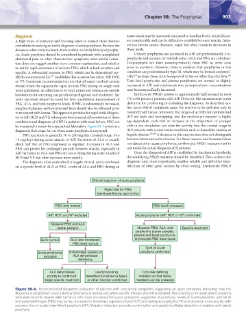
be measured in serum by a specialized laboratory. Figure 58–6 presents a cells in the circulation can raise the activity into the normal range in
diagnostic flow chart for use when acute porphyria is suspected. AIP patients with a concurrent condition such as hemolytic anemia or
PBG excretion is generally 50 to 200 mg/day (normal range: 0 to hepatic disease. 229,230 A decrease in this enzyme also does not distinguish
~4 mg/day) during acute attacks of AIP. Excretion of ALA is usually between latent and active disease. For these reasons, and because it does
about half that of PBG (expressed as mg/day). Increases in ALA and not detect other acute porphyrias, erythrocyte PBGD measurement in
PBG can persist for prolonged periods between attacks, especially in not useful for initial diagnosis of ill patients.
AIP. Increases in ALA and PBG are less striking during acute attacks of Once the diagnosis of AIP is established by biochemical methods,
HCP and VP and often decrease more rapidly. the underlying PBGD mutation should be identified. This confirms the
The diagnosis of an acute attack is largely clinical, and is not based diagnosis and, most importantly, enables reliable and definitive iden-
on a specific level of ALA or PBG. Levels of ALA and PBG during an tification of other gene carriers by DNA testing. Erythrocyte PBGD
Clinical suspicion of acute porphyria
Rapid test for PBG
(semiquantitative, spot urine)
PBG level normal PBG level increased
AIP, HCP, and VP excluded Acute porphyria (AIP, HCP, or VP) confirmed
Measure PBG and ALA
(same sample) Measure PBG, ALA, and Specific treatment
porphyrins (same sample),
plasma and fecal porphyrins,
Both normal ALA level increased, erythrocyte PBG deaminase
PBG level normal
Type of acute
Acute porphyrlas Differentiate causes of porphyria confirmed
excluded ALA dehydratase
deficiency
ALA dehydratase Lead poisoning, Consider defining
porphyria confirmed: hereditary tyrosinemia type I, mutation so that family
begin specific treatment or other disorder confirmed members can be screened
Figure 58–6. Recommended laboratory evaluation of patients with concurrent symptoms suggesting an acute porphyria, indicating how the
diagnosis is established or excluded by biochemical testing and when specific therapy should be initiated. This schema is not applicable to patients
who were recently treated with hemin or who have recovered from past symptoms suggestive of porphyria. Levels of δ-aminolevulinic acid (ALA)
and porphobilinogen (PBG) may be less increased in hereditary coproporphyria (HCP) and variegate porphyria (VP) and decrease more quickly with
recovery than in acute intermittent porphyria (AIP). Mutation detection provides confirmation and greatly facilitates detection of relatives with latent
porphyria.
Kaushansky_chapter 58_p0889-0914.indd 903 9/18/15 5:58 PM