
Page 923 - Williams Hematology ( PDFDrive )
P. 923
898 Part VI: The Erythrocyte Chapter 58: The Porphyrias 899
EPP was usually described in the past as an autosomal dominant immunoglobulin, complement, and periodic acid-Schiff–positive
disorder, with variable penetrance. However, it was noted that EPP mucopolysaccharides. Basement membrane abnormalities are less
121
patients have only 30 percent or less of normal FECH activity, rather than marked than in other forms of porphyria. 122
50 percent, which would be expected in an autosomal dominant condi- Protoporphyric hepatopathy is a feared complication that develops
tion. It was then shown that, in addition to a severe FECH mutation, in less than 5 percent of patients, and is attributed to the cholestatic
a low-expression (hypomorphic) intronic polymorphism (a –23C→T effects of excess protoporphyrin presented to the liver. This complica-
transition) is found in the other FECH allele of patients with EPP, which tion may begin with chronic abnormalities in liver function tests and
is inherited from the other parent. 107–109 This transition favors the use then progress rapidly as a vicious cycle of increasing protoporphyrin
of a cryptic acceptor splice site 63 bases upstream of the normal splice levels in plasma and erythrocytes and worsening liver function and pho-
site. The aberrantly spliced mRNA contains a premature stop codon and tosensitivity. Hepatopathy is sometimes precipitated by another cause
109
is degraded by a nonsense-mediated decay mechanism. The result is of liver dysfunction such as viral or alcoholic hepatitis. Protoporphyrin
a lower steady-state level of wild-type FECH mRNA. Coinheritance is cholestatic, and can form crystalline structures in hepatocytes and
of the hypomorphic allele in trans to a loss-of-function mutant allele impair mitochondrial function, leading to decreased hepatic bile forma-
110
was found in 98 percent of French cases with EPP, and with a similar tion and flow. 123,124 Accumulated protoporphyrin may appear as brown
105
frequency in South African patients. The frequency of the IVS3–48C pigment in hepatocytes, Kupffer cells, and biliary canaliculi, and these
hypomorphic allele is common in the white population, and by itself has deposits are doubly refractive with a Maltese cross appearance under
125
no phenotype. Its frequency varies widely in different populations and polarizing microscopy. DNA microarray studies in explanted livers of
relates to the observed differences in the prevalence of EPP. 103–105 patients with hepatopathy revealed significant changes in expression of
Other underlying genetic mechanisms must be considered in newly several genes involved in wound-healing, organic anion transport, and
identified EPP families. In a few families, a severe FECH mutation, at oxidative stress. 126
least one of which must produce some FECH enzyme, is inherited from
each parent and the hypomorphic allele is not present. Interestingly, Clinical Features
EPP in such families is sometimes associated with seasonal palmar ker- Photosensitivity is present from early childhood in almost all cases.
atoderma, unusual neurologic symptoms, less-than-expected increases Parents may observe that an affected infant cries and develops skin
in erythrocyte protoporphyrin and absence of liver dysfunction. 111 swelling and erythema when exposed to sunlight. Although EPP is the
XLP was first perceived as a variant form of EPP in which FECH most common porphyria in children, there is often considerable delay
mutations were absent. After family studies suggested sex-linked inher- in diagnosis.
itance, gain-of-function mutations of ALAS2 (the only heme pathway Cutaneous photosensitivity in EPP is acute and nonblistering,
enzyme found on the X chromosome) were discovered. This is the which is distinctly different from the more chronic, blistering skin
37
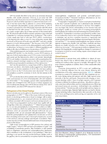
only porphyria caused by mutations of ALAS, the first enzyme in the manifestations of the other cutaneous porphyrias. Table 58–3 tabulates
pathway. symptoms in a series of 32 patients with EPP. Skin symptoms are usu-
EPP can develop late in life in patients with clonal hematologic dis- ally worse during spring and summer and affect light-exposed areas,
orders and expansion of a clone of hematopoietic cells with mutations especially of the face and hands. Characteristically, stinging or burn-
of a FECH allele. 112,113 For example, a patient with a myeloproliferative ing pain develops within 1 hour of sunlight exposure, and if exposure
disorder later developed severe EPP because of clonal expansion of a continues is followed by erythema and edema—described as solar urti-
cell of erythropoietic lineage with a FECH deletion and the IVS3–48C/T caria, sometimes with petechiae, and less commonly purpura. Blistering
polymorphism, and died of EPP-induced liver disease. 114 and crusted lesions are uncommon. Artificial lights may contribute to
127
photosensitivity. Patients typically avoid sunlight and may display no
objective cutaneous signs. Repeated light exposure can lead to chronic
Pathogenesis of the Clinical Findings changes including leathery hyperkeratotic skin especially on the dorsa
Marrow reticulocytes are thought to be the primary source of the excess of the hands and finger joints, mild scarring, and separation of the nail
protoporphyrin in EPP. 115,116 Most of the excess erythrocyte protopor- plate (onycholysis).
phyrin in circulating erythrocytes is found in younger cells as metal-
free protoporphyrin (i.e., not complexed with zinc), in contrast to other
conditions associated with increased erythrocyte protoporphyrin con- TABLE 58–3. Common Clinical Features of Erythropoietic
tent. Metal-free protoporphyrin declines much more rapidly with red Protoporphyria from a Series of 32 Cases
cell age than it does zinc protoporphyrin. 115,116 Metal-free protoporphy- Symptoms and Signs Incidence (% of Total)
rin, but not zinc protoporphyrin, is released from erythrocytes follow-
ing solar irradiation, which may explain why lead intoxication and iron Burning 97
deficiency, which are associated with elevated erythrocyte zinc pro- Edema 94
toporphyrin levels, are not associated with photosensitivity. Excess Itching 88
117
metal-free protoporphyrin enters plasma from reticulocytes, as well as
from circulating erythrocytes, and is taken up by hepatocytes, excreted Erythema 69
in bile and feces, and may undergo enterohepatic recirculation. Hepa- Scarring 19
tocytes may also provide a limited additional source of excess protopor- Vesicles 3
phyrin in this disease.
Light-excited protoporphyrin in EPP generates free radicals and Anemia 27
119
singlet oxygen, which in EPP can lead to peroxidation of lipids and Cholelithiasis 12
118
crosslinking of membrane proteins. Skin irradiation in EPP patients Abnormal liver function results 4
leads to complement activation and polymorphonuclear chemotaxis,
which contributes to the development of skin pathology. Skin histo- Data from Bloomer J, Wang Y, Singhal A, et al: Molecular studies of
120
pathology is not specific but may include thickened capillary walls in liver disease in erythropoietic protoporphyria, J Clin Gastroenterol
the papillary dermis surrounded by amorphous hyaline-like deposits, 2005 Apr;39(4 Suppl 2):S167–S175.
Kaushansky_chapter 58_p0889-0914.indd 898 9/18/15 5:58 PM