
Page 998 - Williams Hematology ( PDFDrive )
P. 998
972 Part VII: Neutrophils, Eosinophils, Basophils, and Mast Cells Chapter 63: Basophils, Mast Cells, and Related Disorders 973
of infection with parasites such as Strongyloides, in which a greater than ligand-independent activation of the KIT receptor, was first identified
177
fourfold increase in mast cell numbers can occur. In such settings, in a long-term cell line derived from a patient with MCL. It then was
165
mast cell numbers return toward normal upon resolution of the infec- detected in mononuclear cells in the blood of patients with mastocytosis
178
tion. Finally, mast cell numbers can be increased several-fold in lymph who had an associated hematologic disorder, as a somatic mutation
nodes draining areas of tumor growth 164,166 and in subjects with stem in lesional tissue obtained from one patient with an aggressive form of
179
cell diseases and lymphoproliferative diseases, including lymphoma in mastocytosis and from a second patient with an indolent form of UP,
the marrow and in association with CML. 164,167–169 and in the skin, but not the marrow and blood, of an 11-month-old
child with mastocytosis. 180
Together these findings suggest the mutation occurs initially in a
DISORDERS OF MAST CELLS: mast cell progenitor and that, as the clone expands, it becomes detect-
HYPERPLASIA AND NEOPLASIA able in the marrow, blood, and skin lesions. The Asp816Val mutation, or
similar 816 activating mutations that result in the substitution of pheny-
DEFINITION AND HISTORY lalanine or tyrosine for aspartate, now are believed to occur in more than
181
A group of systemic disorders associated with significant increases in mast 90 percent of adult patients with mastocytosis. Mutations at codon
cell numbers in the skin and internal organs have been brought together 816 have been identified in a subset of pediatric patients, whereas other
under the term mastocytosis. The first report of a primary mast cell dis- pediatric patients exhibit KIT mutations elsewhere, including within
order is attributed to Unna who, in 1887, reported that the skin lesions the extracellular domain. These other mutations also cause constitutive
170
of urticaria pigmentosa (UP) 171,172 contained numerous mast cells. Ellis activation in KIT to a varying degree. 181
173
recognized the systemic nature of the disorder in 1949. In addition to the The extent to which the presence of various KIT mutations, and the
systemic disorders classified as mastocytosis, localized cutaneous aggre- anatomical distribution of the affected cells, can be used to predict prog-
gates of mast cells, ranging from mast cell nevi and mastocytomas in infants nosis or disease severity in patients with mastocytosis remains under
and children to multiple nodules in older children, may occur. 174,175 investigation. Moreover, additional “gain-of-function” mutations of KIT
The clinical pattern of disease in mastocytosis and its prognosis in human subjects with mastocytosis have been reported. For example,
can vary substantially among patients (see “Course and Prognosis” a novel form of mastocytosis with a KIT mutation in the transmem-
182
below). A consensus classification for mastocytosis has been developed brane domain (Phe522Cys) has been described. In a second example,
to address the issue and to provide guidelines regarding prognosis and a PRKG2-PDGFRB fusion was identified in a patient presenting with
183
treatment (Table 63–5). Patients with indolent disease, who compose increased numbers of mast cells and peripheral basophilia. The lat-
176
the great majority of subjects with mastocytosis, can expect a normal ter case falls within the World Health Organization category of myeloid
life span. Patients with systemic mastocytosis with associated clonal, neoplasms with PDGFRB rearrangements, rather than being catego-
hematologic non–mast-cell-lineage disease (SM-AHNMD) have a rized as a subvariant of mastocytosis. Gain-of-function mutations of
184
prognosis determined by the associated hematologic disorder. Patients KIT also have been reported in gastrointestinal stromal tumors. Addi-
with aggressive systemic mastocytosis (ASM) generally have a 3- to tional genetic lesions have been reported in aggressive mastocytosis and
5-year survival. Mast cell leukemia (MCL) is often rapidly fatal. in patients with SM-AHNMD, including mutations in JAK2, TET2,
NRAS, and KRAS. 185
ETIOLOGY AND PATHOGENESIS
Activating mutations in KIT, which encodes the SCF receptor, a mem- CLINICAL FEATURES
ber of the type III receptor tyrosine kinase family, have been docu- The organs most frequently involved in systemic mastocytosis are the
mented in patients with mastocytosis. Several lines of evidence indicate skin, lymph nodes, liver, spleen, marrow, and gastrointestinal tract.
such mutations can be involved in the pathogenesis of the disease.
The most common of these mutations (Asp816Val), which results in The Skin
The usual presenting lesion of mastocytosis in the skin is UP/maculopap-
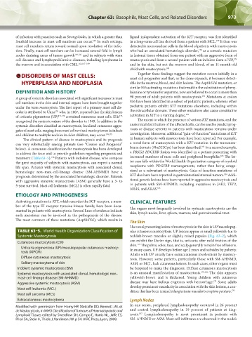
TABLE 63–5. World Health Organization Classification of ular cutaneous mastocytosis. UP lesions appear as small yellowish-tan to
Systemic Mastocytosis reddish-brown macules or slightly raised papules (Fig. 63–2), which
Cutaneous mastocytosis (CM) can exhibit the Darier sign, that is, urticaria after mild friction of the
174
Urticaria pigmentosa (UP)/maculopapular cutaneous mastocy- skin. The palms, soles, face, and scalp generally remain free of lesions.
In many cases, UP develops before age 2 years and subsides by puberty.
tosis (MPCM) Adults with UP usually have extracutaneous involvement by mastocy-
Diffuse cutaneous mastocytosis tosis. However, some patients, particularly those with SM-AHNMD,
Solitary mastocytoma of skin ASM, or MCL, lack cutaneous lesions. In such cases, other organs must
Indolent systemic mastocytosis (ISM) be biopsied to make the diagnosis. Diffuse cutaneous mastocytosis
Systemic mastocytosis with associated clonal, hematologic non– is an unusual manifestation of mastocytosis. 175,186 The skin appears
mast-cell-lineage disease (SM-AHNMD) yellowish-brown and is thickened. Young children with cutaneous
175
Aggressive systemic mastocytosis (ASM) disease may have bullous eruptions with hemorrhage. Some adults
Mast cell leukemia (MCL) develop prominent vascularity in association with the skin lesions, a con-
dition that has been termed telangiectasia macularis eruptiva perstans.
175
Mast cell sarcoma (MCS)
Extracutaneous mastocytoma Lymph Nodes
Modified with permission from Horny HP, Metcalfe DD, Bennett JM, et In one series, peripheral lymphadenopathy occurred in 26 percent
al: Mastocytosis, in WHO Classification of Tumours of Haematopoietic and and central lymphadenopathy in 19 percent of patients at diag-
187
Lymphoid Tissues edited by Swerdlow SH, Campo E, Harris NL, Jaffe ES, nosis. Lymphadenopathy is most prominent in patients with
Pileri SA, Stein H, Thiele J, Vardiman JW. p 54. IARC Press, Lyon, 2008. SM-AHNMD or ASM. Mast cell infiltrates are observed in the node’s
Kaushansky_chapter 63_p0965-0982.indd 973 9/18/15 11:01 PM