
Page 999 - Williams Hematology ( PDFDrive )
P. 999
974 Part VII: Neutrophils, Eosinophils, Basophils, and Mast Cells Chapter 63: Basophils, Mast Cells, and Related Disorders 975
Spleen
Splenic involvement at diagnosis has been reported in approximately
half of patients with systemic disease. 187,189 Mast cells most commonly
occurred in a paratrabecular distribution, followed by perifollicu-
lar, follicular, and diffuse infiltrates. Trabecular and capsular fibrosis
and eosinophilic infiltration also were observed, and extramedullary
hematopoiesis was present in most cases. On H&E stained sections,
the infiltrates of mast cells produced lesions that may resemble those
of T-cell lymphoma, follicular hyperplasia, follicular lymphoma, mye-
loproliferative neoplasms, hairy cell leukemia, or a granulomatous pro-
cess. Splenomegaly has also been reported in the absence of infiltration
of the spleen by mast cells. Increased splenic weights greater than
190
700 g generally occurred in patients within unfavorable categories of
mastocytosis.
Marrow
The majority of adults with systemic mast cell disease have focal mast
cell lesions in the marrow, 189,191–194 typically appearing as foci of spin-
dle-shaped mast cells in a fibrotic background (Fig. 63–3), sometimes
with associated eosinophils and T and B lymphocytes. These focal mast
cell lesions are the major criterion in the diagnosis of systemic mastocy-
tosis (Table 63–6). Reticulin staining may be increased, and Masson
176
trichome staining may reveal collagen deposition. In specimens exten-
sively involved by mast cell lesions, the bony trabeculae may be moder-
ately to markedly thickened. Aggressive variants of mastocytosis, such
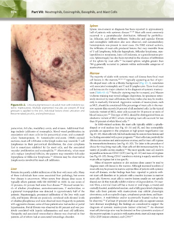
Figure 63–2. Urticaria pigmentosa in an adult man with indolent sys- as MCL, should be considered if the percentage of mast cells in the mar-
temic mastocytosis. Multiple pigmented macules are present. If local row aspirate film exceeds 20 percent of all nucleated cells. In the typical
pressure is applied to the skin, individual lesions show urtication and leukemic variant of MCL, mast cells account for 10 percent or more of
become raised, pruritic, and erythematous. blood leukocytes. This type of MCL should be distinguished from an
176
aleukemic variant of MCL where circulating mast cells account for less
than 10 percent of white blood cells. 195
In H&E–stained sections, the mast cells typically exhibit a spindle-
paracortex, follicles, medullary cords, and sinuses. Additional find- shaped or oval nucleus (see Fig. 63–3A and B), and fine eosinophilic
ings include infiltrates of eosinophils, blood vessel proliferation in granules are apparent in the cytoplasm at high-power magnification (see
association with mast cells in the paracortical areas, and extramed- Fig. 63–3B). Mast cells with bilobed nuclei may be seen in these lesions and
189
ullary hematopoiesis. In hematoxylin-and-eosin (H&E)–stained is a finding associated with a poor prognosis. Mast cells stain positively for
sections, mast cell infiltrates in the lymph nodes may resemble T-cell chloracetate esterase and aminocaproate esterase, and for mast cell tryptase
lymphomas in their pericortical distribution, the clear cytoplasm by immunohistochemistry (see Fig. 63–3D). The latter is the procedure of
that is sometimes exhibited by the mast cells, and the associated choice for visualizing mast cells. Mast cells exhibit immunoreactivity for a
196
187
vascular proliferation and eosinophilia. Alternatively, when mast variety of paraffin section markers. The more specific mast cell markers
cells replace lymphoid follicles, the pattern may resemble follicular in paraffin sections are CD117 (KIT) (see Fig. 63–3C) and mast cell tryptase
187
hyperplasia or follicular lymphoma. Fibrosis may be observed in (see Fig. 63–3D). Strong CD117 membrane staining is equally sensitive for
lymph nodes involved by mast cell infiltrates. mast cells as tryptase but is less specific.
Films of marrow aspirates or clot sections alone cannot be used to
diagnose mast cell disease in the marrow. Although increased numbers of
Liver mast cells may be present in marrow aspirate films of patients with systemic
Patients frequently exhibit infiltration of the liver with mast cells. Many mast cell diseases, similar findings have been reported in patients with-
of these individuals have some associated liver pathology, but severe out mast cell disorders or in patients with a reactive increase in marrow
liver disease is uncommon. When severe liver disease does occur, it mast cells. However, mast cells in reactive lesions usually are not spindle
typically affects patients with SM-AHNMD or ASM. In one series of shaped, nor do they typically exhibit evidence of degranulation. On mar-
41 patients, 61 percent had some liver disease. Elevated serum lev- row films, a normal mast cell has a round or oval shape, a round and
188
els of alkaline phosphatase, aminotransaminases, 5′-nucleotidase, or centrally located, nonlobated nucleus, and a fully granulated cytoplasm.
γ-glutamyl transpeptidase was detected in approximately half of the Mast cells from patients with mastocytosis may exhibit phenotypic
patients. Hepatomegaly, prominent infiltration of the liver with mast aberrations, such as a spindle shape, cytoplasmic projections, and hypo-
cells, and hepatic fibrosis are positively correlated with elevated levels granulation. A multilobular and/or eccentrically located nucleus may
176
of alkaline phosphatase and were observed more frequently in patients be observed. If at least 25 percent of all mast cells on aspirate smears
with aggressive disease; some of these patients also had ascites or portal have aberrant morphology, the findings are considered to support the
176
hypertension. Portal fibrosis was observed in 68 percent and was pos- diagnosis of systemic mastocytosis (minor criterion). An aberrant
itively correlated with hepatic inflammation and mast cell infiltrates. mast cell phenotype also may be detected on flow cytometric analysis of
Venopathy and associated venoocclusive disease was observed in four the marrow aspirate. In patients with mastocytosis, mast cells may express
patients, all of whom had an associated hematologic disorder. CD2, CD25 (minor criterion), and CD33. 197
Kaushansky_chapter 63_p0965-0982.indd 974 9/18/15 11:01 PM