
Page 1105 - Clinical Immunology_ Principles and Practice ( PDFDrive )
P. 1105
ChaPTEr 79 Lymphomas 1069
This entity usually presents with low clinical stage and large or (q32;q21), t(3;14)(q27;q32), and t(3;14)(p14.1;q32); these
localized inguinal tumors. 17 abnormalities are observed with variable frequency, often depend-
19
FL is indolent but is still incurable with available therapeutic ing on the anatomical site. Although several genes are involved
modalities. The occurrence of BCL2 translocation at a very in these translocations, at least three of them—t(11;18), t(1;14),
early stage of B-cell development might be contributory and t(14;18)—share a common pathway, leading to the activation
20
to the difficulty of eradicating the neoplastic clone with chemo- of NFκB and its downstream targets. By genome-wide DNA
therapy. Clinical parameters have been used to develop prognostic profiling integrated with GEP, differences were detected among
indexes (e.g., the Follicular Lymphoma International Prognostic the three main types of marginal zone lymphomas, lending
Index, adjusted from the International Prognostic Index for support to the current WHO classification that separates these
18
aggressive B-cell lymphoma). Recently, much has also been learned three entities. The translocation t(11;18)(q21;q21) is associated
about the mutational landscape of FL. Mutations in CREBBP, exclusively with low-grade extranodal MALT and is not detected
MLL2, and EZH2 have been shown as extremely common in cases with concurrent low-grade and high-grade tumors, or
18
early events and may be potential therapeutic targets. The in primary extranodal large-cell lymphomas—raising doubts
natural history of the disease is associated with histological whether these primary extranodal lymphomas are, in fact, related
progression at both cellular composition and overall pattern to low-grade MALT. The term extranodal marginal zone lymphoma
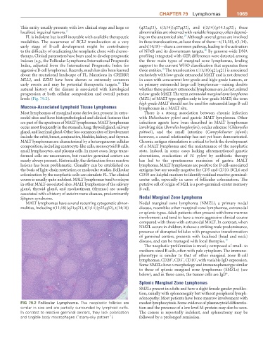
levels (Fig. 79.2). (MZL) of MALT type applies only to low-grade MALT; the term
high-grade MALT should not be used for extranodal large B-cell
Mucosa-Associated Lymphoid Tissue Lymphomas lymphomas in a MALT site.
Most lymphomas of marginal zone derivation present in extra- There is a strong association between chronic infection
nodal sites and have histopathological and clinical features that with Helicobacter pylori and gastric MALT lymphoma. Other
are part of the spectrum of MALT lymphomas. MALT lymphomas infectious agents have been described in MALT lymphomas
occur most frequently in the stomach, lung, thyroid gland, salivary involving skin (Borrelia burgdorferi), ocular adnexae (Chlamydia
gland, and lacrimal gland. Other less common sites of involvement psittaci), and the small intestine (Campylobacter jejuni);
include the orbit, breast, conjunctiva, bladder, kidney, and thymus. however, a causal relationship has not yet been demonstrated.
MALT lymphomas are characterized by a heterogeneous cellular Chronic antigen stimulation is critical to both the development
composition, including centrocyte-like cells, monocytoid B cells, of a MALT lymphoma and the maintenance of the neoplastic
small lymphocytes, and plasma cells. In most cases, large trans- state. Indeed, in some cases lacking aforementioned genetic
formed cells are uncommon, but reactive germinal centers are aberrations, eradication of H. pylori by antibiotic therapy
nearly always present. Historically, the distinction from reactive has led to the spontaneous remission of gastric MALT
lesions has been problematic. Clonality can be established on lymphoma. MALT lymphomas are positive for B cell–associated
the basis of light-chain restriction or molecular studies. Follicular antigens but are usually negative for CD5 and CD10. BCL6 and
colonization by the neoplastic cells can simulate FL. The clinical CD10 are helpful markers to identify residual reactive germinal-
course is usually quite indolent. MALT lymphomas tend to relapse center cells, especially in cases of follicular colonization. The
in other MALT-associated sites. MALT lymphomas of the salivary putative cell of origin of MZL is a post–germinal-center memory
gland, thyroid gland, and mediastinum (thymus) are usually B cell.
associated with a history of autoimmune diseases, predominantly
Sjögren syndrome. Nodal Marginal Zone Lymphoma
MALT lymphomas have several recurring cytogenetic abnor- Nodal marginal zone lymphoma (NMZL), a primary nodal
malities, including t(11;18)(q21;q21), t(1;14)(p22;q32), t(14;18) disease, resembles other marginal zone lymphomas, extranodal
or splenic types. Adult patients often present with bone marrow
involvement and tend to have a more aggressive clinical course
compared with those with extranodal MALT. In contrast, when
NMZL occurs in children, it shows a striking male predominance,
presence of disrupted follicles with progressive transformation
of germinal centers, presents with localized (head and neck)
disease, and can be managed with local therapies. 19
The neoplastic proliferation is mostly composed of small- to
medium-sized B cells, often with pale cytoplasm. The immuno-
phenotype is similar to that of other marginal zone B-cell
−
−
+
lymphomas, CD20 , CD5 , CD10 , with variable IgD expression.
Some NMZLs have a morphology and immunophenotype similar
to those of splenic marginal zone lymphomas (SMZLs) (see
+
below), and in these cases, the tumor cells are IgD .
Splenic Marginal Zone Lymphomas
SMZLs present in adults and have a slight female gender predilec-
tion, usually with splenomegaly but without peripheral lymph-
adenopathy. Most patients have bone marrow involvement with
FIG 79.2 Follicular Lymphoma. The neoplastic follicles are modest lymphocytosis. Some evidence of plasmacytoid differentia-
similar in size and are partially surrounded by lymphoid cuffs. tion and the presence of a low level M-protein may also be seen.
In contrast to reactive germinal centers, they lack polarization The course is reportedly indolent, and splenectomy may be
and tingible body macrophages (“starry-sky pattern”). followed by a prolonged remission.