
Page 449 - Clinical Immunology_ Principles and Practice ( PDFDrive )
P. 449
428 ParT THrEE Host Defenses to Infectious Agents
Leishmania promastigotes gain the capacity to produce IFN-γ, and then migrate to the site
of infection. Thus T cm cells act as a reserve of antigen-reactive
NK T cells that can expand and become effector T cells in response
IL-12 to secondary antigenic challenge.
DC
The generation of RNI by activated macrophages is the primary
IFN-γ mechanism of parasite killing in the murine model. Although
IL-12 IFN-γ-induced production of NO may not be detectable in human
IFN-γ IFN-γ macrophages, inhibition of nitric oxide synthase 2 (NOS2) was
IFN-γ shown to impair killing of intracellular Leishmania.
IFN-γ Th1 Several adaptive immune mechanisms promote parasite
15
IL-4 replication and disease. The progression of murine L. major
IL-10 IFN-γ infection has been correlated with the expansion of Th2 cells
Th2
IL-10 TNF-α and the production of IL-4, IL-5, and IL-10. In susceptible mice,
TGF-β IL-4 production within the first day of infection was shown to
IL-4 IL-10
IL-10 Activation TGF-β downregulate IL-12 receptor β-chain expression and drive the
RNI response to a Th2 phenotype. However, other nonsusceptible
ROI mouse strains appear to be able to overcome an early IL-4 response
IL-10
TGF-β Parasite killing and develop a resistant phenotype, and susceptibility to some
PGE 2 Deactivation L. major strains is not strictly mediated by IL-4 (IL-13 and/or
IL-10 may have a prominent role). The cytotoxic activity of CD8
Parasite Macrophage T cells may promote cutaneous inflammation and lesion pathol-
14
replication ogy. The macrophage production of immune suppressive
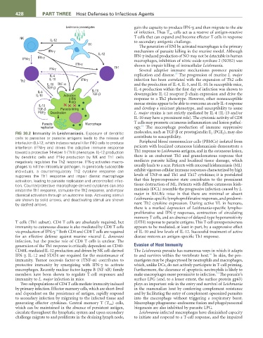
FIG 30.2 Immunity in Leishmaniasis. Exposure of dendritic molecules, such as TGF-β or prostaglandin E 2 (PGE 2 ), may also
cells to parasites or parasite antigens leads to the release of contribute to susceptibility.
interleukin (IL)-12, which induces natural killer (NK) cells to produce Peripheral blood mononuclear cells (PBMCs) isolated from
interferon (IFN)-γ and drives the adaptive immune response patients with localized cutaneous leishmaniasis demonstrate a
toward a protective T-helper 1 (Th1) phenotype. IL-12 production Th1 response to Leishmania antigens, and in the cutaneous lesion,
by dendritic cells and IFN-γ production by NK and Th1 cells there is an exuberant Th1 and granulomatous response that
negatively regulates the Th2 response. IFN-γ activates macro- mediates parasite killing and localized tissue damage, which
phages to kill the intracellular pathogen. In genetically susceptible usually leads to a scar. Patients with mucosal leishmaniasis (ML)
individuals, a counterregulatory Th2 cytokine response can exhibit vigorous cellular immune responses characterized by high
suppress the Th1 response and impair classic macrophage levels of TNF-α and Th1 and Th17 cytokines; it is postulated
activation, leading to parasite replication and uncontrolled infec- that this hyperresponsive state contributes to the prominent
tion. Counterprotective macrophage-derived cytokines can also tissue destruction of ML. Patients with diffuse cutaneous leish-
inhibit the Th1 response, stimulate the Th2 response, and impair maniasis (DCL) resemble the progressive infection caused by L.
classical activation through an autocrine loop. Activating stimuli major in BALB/c mice in that there are minimal or absent
are shown by solid arrows, and deactivating stimuli are shown Leishmania-specific lymphoproliferative responses, and predomi-
by dashed arrows. nant Th2 cytokine expression. During active VL in humans,
there is a marked depression of Leishmania-specific lympho-
proliferative and IFN-γ responses, contraction of circulating
memory T cells, and an absence of delayed-type hypersensitivity
T cells (Th1 subset). CD4 T cells are absolutely required, but (DTH) response to parasite antigens. This T-cell unresponsiveness
immunity to cutaneous disease is also mediated by CD8 T cells appears to be mediated, at least in part, by a suppressive effect
14
via production of IFN-γ. Both CD4 and CD8 T cells are required of IL-10 and low levels of IL-12. Successful treatment of active
for an effective defense against murine visceral L. donovani disease restores an antigen-specific Th1 response.
infection, but the precise role of CD8 T cells is unclear. The
generation of the Th1 response is critically dependent on CD40- Evasion of Host Immunity
CD40L-mediated IL-12 production and driven by NK cell–derived The Leishmania parasite has numerous ways in which it adapts
16
IFN-γ. IL-12 and STAT4 are required for the maintenance of to and survives within the vertebrate host. In skin, the pro-
immunity. Tumor necrosis factor-α (TNF-α) contributes to mastigotes may be phagocytosed by neutrophils and macrophages,
protective immunity by synergizing with IFN-γ to activate which, unlike DCs, do not actively participate in T-cell priming.
macrophages. Recently nuclear factor-kappa B (NF-κB) family Furthermore, the clearance of apoptotic neutrophils is likely to
13
members have been shown to regulate T-cell responses and make macrophages more permissive to infection. The parasite’s
immunity to L. major infection in mice. surface LPG (and, to a lesser extent, the surface protein gp63)
Two subpopulations of CD4 T cells mediate immunity induced plays an important role in the entry and survival of Leishmania
by primary infection. Effector memory cells, which are short-lived in the mammalian host by conferring complement resistance
and dependent on the persistence of antigen, rapidly respond and by facilitating the entry of complement-opsonized parasites
to secondary infection by migrating to the infected tissue and into the macrophage without triggering a respiratory burst.
generating effector cytokines. Central memory T (T cm ) cells, Macrophage phagosome–endosome fusion and phagolysosomal
which can be maintained in the absence of persistent antigen, biogenesis are also inhibited by parasite LPG.
circulate throughout the lymphatic system and upon secondary Leishmania-infected macrophages have diminished capacity
challenge migrate to and proliferate in the draining lymph node, to initiate and respond to a T-cell response, and the impaired