
Page 453 - Clinical Immunology_ Principles and Practice ( PDFDrive )
P. 453
432 ParT THrEE Host Defenses to Infectious Agents
Pathogen Innate immune response Intestinal response
Trophozoite
Complement Villous atrophy and
Giardia Inflammatory crypt hyperplasia
Macrophage mediators
IL-6, IL-8, IL-1
Neutrophil Epithelial
Sporozoite GM-CSF, GROα, damage
Eosinophil prostaglandins
Cryptosporidium
NK cell ROI, RNI
Trophozoite
Proteases
Erosions and
Enterocyte ulcerations
Entamoeba (Cryptosporidium)
Cytokines
B- and T-lymphocyte activation Secretion
Malabsorption
Acquired immune response
Exudation
Host protection Diarrhea
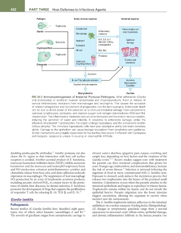
FIG 30.3 Immunopathogenesis of Intestinal Protozoal Pathogens. After adherence (Giardia
and Entamoeba) or epithelial invasion (Entamoeba and Cryptosporidium), there is release of
various inflammatory mediators from macrophages and neutrophils. This causes the activation
of resident phagocytes and recruitment of phagocytes into the lamina propria. Enterocyte death
can be due to direct action of the parasites or to immune-mediated damage from complement,
cytotoxic lymphocytes, proteases, and reactive oxygen and nitrogen intermediates (ROI and RNI,
respectively). The inflammatory mediators also act on enterocytes and the enteric nervous system,
inducing the secretion of water and chloride. In response to enterocyte damage, under the
influence of activated T lymphocytes, the crypts undergo hyperplasia, and the villi become shorter
(villous atrophy). The immature hyperplastic cells have poor absorptive ability but retain secretory
ability. Damage to the epithelium can cause leakage (exudation) from lymphatics and capillaries.
Similar mechanisms are probably responsible for the diarrhea that occurs in infection with Cyclospora
and Isospora. Isospora is unique in causing an eosinophilic infiltrate.
27
shedding ameba-specific antibodies. Amebic proteases can also chronic watery diarrhea, epigastric pain, nausea, vomiting, and
cleave the Fc region so that interaction with host cell surface weight loss, depending on host factors and the virulence of the
receptors is avoided. Another secreted product of E. histolytica, Giardia strain. 30,31 Recent studies suggest even with treatment
monocyte locomotion inhibition factor (MLIF), inhibits monocyte the parasite can elicit intestinal complications that persist for
locomotion and the monocyte and neutrophil respiratory burst years. Younger age, malnutrition, and immunodeficiency increase
31
and NO production; enhances antiinflammatory cytokine and the risk of severe disease. Infection is initiated following the
chemokine release from host cells; and alters adhesion molecule ingestion of food or water contaminated with G. lamblia cysts.
expression on macrophages. The suppression of host macrophage Exposure to stomach acids induces the excystation process that
NO production by an array of trophozoite secretory products, releases two trophozoites into the lumen of the proximal small
including parasite-derived PGE 2 , is a major factor in the persis- intestine. Colonization occurs when the parasite attaches to the
tence of amebic liver abscesses. In chronic infection, E. histolytica intestinal epithelium and begins to reproduce by binary fission.
promotes the development of Tregs that suppress the proliferation Trophozoites remain within the lumen and do not invade the
of responder T cells by releasing IL-10, TGF-β, and IL-35. 28 epithelial barrier. Parasite migration into the lower intestine
triggers encystation, allowing the organism to survive when
Giardia lamblia excreted into the environment.
The G. lamblia trophozoite initiates adherence to the intestinal
Pathogenesis epithelium via a surface mannose-binding lectin. Histopathologi-
Recent studies of Giardia lamblia have identified eight geno- cal changes in symptomatic giardiasis range from a normal
30
types, two of which infect humans (assemblages A and B). appearance to increased crypt-villous ratios, epithelial damage,
The severity of giardiasis ranges from asymptomatic carriage to and chronic inflammatory infiltrate in the lamina propria (see