
Page 510 - Clinical Immunology_ Principles and Practice ( PDFDrive )
P. 510
490 ParT fOur Immunological Deficiencies
KEY CONCEPTS Regardless of the molecular cause, treatment with corticoster-
Combined Immunodeficiency oids and cyclosporine are effective in controlling inflammation,
improving skin lesions, and reducing lymphadenopathy and
• Arbitrarily defined as a T-cell deficiency with >300–500 autologous hepatosplenomegaly. Ultimately, a cure can be achieved only
CD3 circulating cells/µL, which may be reduced or dysfunctional. with HSCT. 11
• Circulating autologous T cells may be the typical feature of the defect
or may be caused by genetic variants leading to an incomplete defect SEVERE COMBINED IMMUNE DEFICIENCY
(leaky severe combined immunodeficiency [SCID]).
• Clinical manifestations may be indistinguishable from SCID. SCID is characterized by near complete absence of circulating
• Others may present with SCID and aberrant inflammatory syndrome
(Omenn syndrome) consisting of erythroderma, lymphadenopathy, autologous T cells and impaired B-cell function. Defects in the
and eosinophilia. differentiation and maturation of T-lineage cells in the thymus
• Frequently, the predominant presenting manifestation can be autoim- are a primary cause of T-cell deficiency (Fig. 35.1). In particular,
munity or lymphoid malignancy. mutations affecting T-cell activation or downstream signaling
• In vitro responses to mitogens and T-cell receptor excision circle molecules can result in SCID or CID (Fig. 35.2).
(TREC) levels may be depressed or normal. The T-cell repertoire is
often abnormal, as are T-cell subpopulations (regulatory T cells [Tregs] SCID With T-Cell Lymphopenia (T B )
−
+
and memory cells). Immunoglobulins and specific antibodies are − +
variable. This T B group of patients (Table 35.2) is the most frequently
identified phenotype of SCID (28 to 50% of all cases) and is
believed to occur at a frequency of 1:50,000 live births. 1
restricted because of inefficient deletion and suppression of
self-reactive T cells and dysregulated lymphostromal cross-talk γc (IL-2Rγ) Deficiency
in the thymus. The condition is associated with hypomorphic Common gamma chain (γc) or interleukin-2 receptor gamma
mutations of various SCID-causing genes, including RAG1/2, (IL-2Rγ) deficiency (OMIM #300400) is transmitted in an
Artemis, DNA ligase IV, RMRP, ZAP-70, IL2RG, IL7Rα, CD3δ, X-linked manner. Affected males have mutations in the IL-2Rγ
and ADA (Table 35.1). In addition to typical SCID manifestations, chain, also known as the common gamma chain because of its
patients present with features including generalized erythroderma, critical role as a coreceptor in IL-4, IL-7, IL-9, IL-15, and IL-21
which may be accompanied by alopecia, loss of eyebrows and receptors. Multiple pathways downstream of these receptors are
eyelashes. Marked enlargement of lymph nodes, liver, and spleen essential for differentiation and growth of T cells and NK cells,
are frequently present. hence the abnormal immune profile and profound defect in
The hallmarks of laboratory findings are elevated serum T-cell and NK-cell maturation. Affected males typically present
immunoglobulin E (IgE) levels and increased eosinophil counts. at 3–6 months of age, with failure to thrive and repeated microbial
T-cell number may be normal or even elevated, but the T-cell and fungal infections. Infections are fatal unless HSCT is
repertoire is skewed with overrepresentation of a few T-cell clones administered.
9
and underexpression or absence of most other Vβ families Evaluation of the immune system typically reveals extremely
−
− +
(Chapter 8). Skin biopsy reveals acanthosis and parakeratosis. low to absent T and NK cells, while sparing B cells (T B NK ).
Dyskeratosis and spongiosis are seen in the Malpighian layer, In vitro responses to mitogens are profoundly depressed, TRECs
and vacuolization is often observed in the basal layer. The thymus are undetectable, and the thymus appears dysplastic with absence
is dysplastic, and expression of the transcription factor auto- of Hassall corpuscles and disrupted architecture.
immune regulator (AIRE) in medullary thymic epithelial cells γc deficiency may have an atypical presentation because of
is reduced. 10 hypomorphic mutations. Various degrees of autologous T-cell
or NK-cell levels have been reported. Importantly, patients with
the R222C mutation can present with SCID-like symptoms but
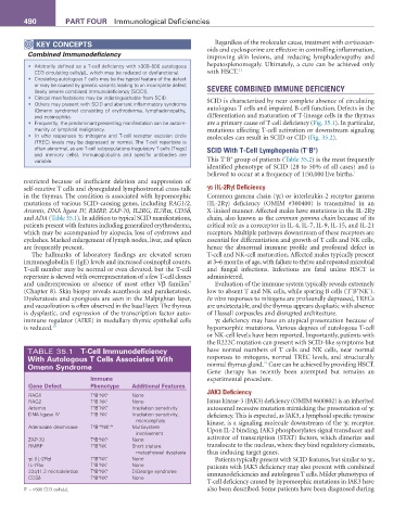
TABLE 35.1 T-Cell Immunodeficiency have normal numbers of T cells and NK cells, near normal
With autologous T Cells associated With responses to mitogens, normal TREC levels, and structurally
12
Omenn Syndrome normal thymus gland. Cure can be achieved by providing HSCT.
Gene therapy has recently been attempted but remains an
Immune experimental procedure.
Gene Defect Phenotype additional features
−
+
RAG1 T B NK + None JAK3 Deficiency
RAG2 T B NK + None Janus kinase-3 (JAK3) deficiency (OMIM #600802) is an inherited
−
+
−
+
Artemis T B NK + Irradiation sensitivity autosomal recessive mutation mimicking the presentation of γc
DNA ligase IV T B NK + Irradiation sensitivity, deficiency. This is expected, as JAK3, a lymphoid specific tyrosine
−
+
microcephaly kinase, is a signaling molecule downstream of the γc receptor.
−/+
+
Adenosine deaminase T B NK −/+ Multisystem Upon IL-2 binding, JAK3 phosphorylates signal transducer and
involvement
ZAP-70 T B NK + None activator of transcription (STAT) factors, which dimerize and
+
+
+
+
RMRP T B NK − Short stature, translocate to the nucleus, where they bind regulatory elements,
metaphyseal dysplasia thus inducing target genes.
+
+
γc (IL-2Rγ) T B NK − None Patients typically present with SCID features, but similar to γc,
IL-7Rα T B NK + None patients with JAK3 deficiency may also present with combined
+
+
+
+
22q11.2 microdeletion T B NK + DiGeorge syndrome immunodeficiencies and autologous T cells. Milder phenotypes of
+
+
CD3δ T B NK + None
T-cell deficiency caused by hypomorphic mutations in JAK3 have
+
T = >500 CD3 cells/µL also been described. Some patients have been diagnosed during