
Page 640 - Clinical Immunology_ Principles and Practice ( PDFDrive )
P. 640
CHaPter 44 Atopic and Contact Dermatitis 615
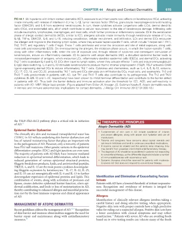
fiG 44.1 (A) In patients with irritant contact dermatitis (ICD), exposure to an irritant exerts toxic effects on keratinocytes (KCs), activating
innate immunity with release of interleukin (IL)-1α, IL-1β, tumor necrosis factor (TNF)-α, granulocyte macrophage–colony-stimulating
factor (GM-CSF), and IL-8 from epidermal keratinocytes. In turn, these cytokines activate Langerhans cells (LCs), dermal dendritic
cells (DCs), and endothelial cells, all of which contribute to cellular recruitment to the site of keratinocyte damage. Infiltrating cells
include neutrophils, lymphocytes, macrophages, and mast cells, which further promote an inflammatory cascade. (B) In the sensitization
phase of allergic contact dermatitis (ACD), similar to ICD, allergens activate innate immunity through keratinocyte release of IL-1α,
IL-1β, TNF-α, GM-CSF, IL-8, and IL-18, inducing vasodilation, cellular recruitment, and infiltration. LCs and dermal DCs encounter
the allergen and migrate to the draining lymph nodes, where they activate hapten-specific T cells, which include T-helper cell-1 (Th1),
Th2, Th17, and regulatory T cells (Tregs). These T cells proliferate and enter the circulation and site of initial exposure, along with
mast cells and eosinophils (EOS). On re-encountering the allergen, the elicitation phase occurs, in which the hapten-specific T cells,
along with other inflammatory cells, enter the site of exposure and, through release of cytokines and consequent stimulation of
keratinocytes, induce an inflammatory cascade. (C) In patients with atopic dermatitis (AD), a disturbed epidermal barrier leads to
increased permeation of antigens, which encounter LCs, inflammatory dendritic epidermal cells (IDECs), and dermal DCs, activating
Th2 T cells to produce IL-4 and IL-13. DCs then travel to lymph nodes, where they activate effector T cells and induce immunoglobulin
E (IgE) class-switching. IL-4 and IL-13 stimulate keratinocytes to produce thymic stromal lymphopoietin (TSLP). TSLP activates OX40
ligand–expressing dermal DCs to induce inflammatory Th2 T cells. Cytokines and chemokines, such as IL-4, IL-5, IL-13, eotaxins,
CCL17, CCL18, and CCL22, produced by Th2 T cells and DCs stimulate skin infiltration by DCs, mast cells, and eosinophils. Th2 and
Th22 T cells predominate in patients with AD, but Th1 and Th17 T cells also contribute to its pathogenesis. The Th2 and Th22
cytokines (IL-4/IL-13 and IL-22, respectively) have been shown to inhibit terminal differentiation and contribute to the barrier defect
in patients with AD. Thus both the barrier defects and immune activation alter the threshold for ICD, ACD, and self-reactivity in
patients with AD. MBP, Major basic protein. (Figure adapted from Gittler JK, Krueger JG, Guttman-Yassky E. Atopic dermatitis results
in intrinsic and immune abnormalities: implications for contact dermatitis. J Allergy Clin Immunol 2013;131:300–13.)
the TSLP–Th2–ILC2 pathway plays a critical role in initiation tHeraPeUtiC PriNCiPLeS
of AD. 11
Atopic Dermatitis (AD)
Epidermal Barrier Dysfunction
• Fundamentals of skin care in AD include avoidance of irritants
The clinically dry skin and increased transepidermal water loss and proven allergens, along with proper skin hydration and use of
(TEWL) in AD reflects underlying skin barrier dysfunction and moisturizers.
loss of natural moisturizing factor that plays an important role • Patients and caregivers have concerns about topical steroids and
in the pathogenesis of AD. However, only a minority of patients calcineurin inhibitors and tend to underuse prescribed medications.
have FLG null mutations. Other genetic variants in the epidermal • If eczema cannot be cleared and the patients keep relapsing, they
may benefit from proactive intermittent antiinflammatory therapy.
differentiation complex (EDC) and tight junctions are even rarer. • The diagnosis of AD should be reconsidered in patients not responding
The majority of patients with AD likely have immune-mediated to conventional therapy, as the differential diagnosis includes a number
reduction in epidermal terminal differentiation, which leads to of immunodeficiencies with eczematous rash.
reduced generation of various epidermal structural proteins, • Systemic therapies should be reserved for patients with moderate-
filaggrin breakdown products, lipids, and antimicrobial peptides. to-severe AD that does not respond to conventional therapy.
TSLP, IL-4, and IL-13 are the most potent cytokines downregulat-
12
ing filaggrin expression by keratinocytes. IL-17, IL-22, IL-25,
and IL-33 can act synergistically with IL-4 and IL-13 to further
downregulate expression of epidermal proteins and lipids. This Identification and Elimination of Exacerbating Factors
combination of events, along with activation of proteases and Irritants
lipases, creates defective epidermal barrier function, alters epi- Patients with AD have a lowered threshold of irritant responsive-
dermal acidification, and leads to loss of moisturization in AD, ness. Recognition and avoidance of irritants is integral for
thereby contributing to enhanced allergen and microbial penetra- successful management of this disease.
tion met by the host immune response and clinical appearance
of AD. Allergens
Identification of clinically relevant allergens involves taking a
MANAGEMENT OF ATOPIC DERMATITIS careful history and doing selective testing, when appropriate.
Negative skin tests with proper controls have a high predictive
Recent guidelines address the management of AD. 13,14 Recognition value for ruling out a suspected allergen. Positive skin tests have
of skin barrier and immune abnormalities suggests the need for a lower correlation with clinical symptoms and may reflect
16
barrier repair and maintenance along with antiinflammatory sensitization. Patients with severe AD who are avoiding foods
measures. 15 based on in vitro testing results can tolerate many of the foods