
Page 713 - Clinical Immunology_ Principles and Practice ( PDFDrive )
P. 713
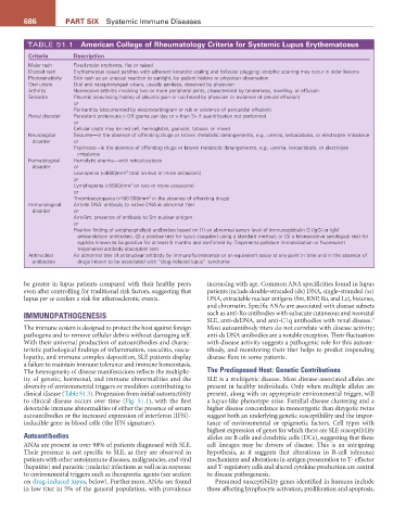
686          Part SIX  Systemic Immune Diseases


         TABLE 51.1  american College of rheumatology Criteria for Systemic Lupus Erythematosus
          Criteria     Description
          Malar rash   Fixed malar erythema, flat or raised
          Discoid rash  Erythematous raised patches with adherent keratotic scaling and follicular plugging; atrophic scarring may occur in older lesions
          Photosensitivity  Skin rash as an unusual reaction to sunlight, by patient history or physician observation
          Oral ulcers  Oral and nasopharyngeal ulcers, usually painless, observed by physician
          Arthritis    Nonerosive arthritis involving two or more peripheral joints, characterized by tenderness, swelling, or effusion
          Serositis    Pleuritis (convincing history of pleuritic pain or rub heard by physician or evidence of pleural effusion)
                       or
                       Pericarditis (documented by electrocardiogram or rub or evidence of pericardial effusion)
          Renal disorder  Persistent proteinuria > 0.5 grams per day or > than 3+ if quantification not performed
                       or
                       Cellular casts may be red cell, hemoglobin, granular, tubular, or mixed
          Neurological   Seizures—in the absence of offending drugs or known metabolic derangements, e.g., uremia, ketoacidosis, or electrolyte imbalance
           disorder    or
                       Psychosis—in the absence of offending drugs or known metabolic derangements, e.g., uremia, ketoacidosis, or electrolyte
                        imbalance
          Hematological   Hemolytic anemia—with reticulocytosis
           disorder    or
                       Leukopenia (<4000/mm  total on two or more occasions)
                                       3
                       or
                       Lymphopenia (<1500/mm  on two or more occasions)
                                        3
                       or
                       Thrombocytopenia (<100 000/mm  in the absence of offending drugs)
                                              3
          Immunological   Anti-ds DNA: antibody to native DNA in abnormal titer
           disorder    or
                       Anti-Sm: presence of antibody to Sm nuclear antigen
                       or
                       Positive finding of antiphospholipid antibodies based on (1) an abnormal serum level of immunoglobulin G (IgG) or IgM
                        anticardiolipin antibodies; (2) a positive test for lupus coagulant using a standard method; or (3) a false-positive serological test for
                        syphilis known to be positive for at least 6 months and confirmed by Treponema pallidum immobilization or fluorescent
                        treponemal antibody absorption test
          Antinuclear   An abnormal titer of antinuclear antibody by immunofluorescence or an equivalent assay at any point in time and in the absence of
           antibodies   drugs known to be associated with “drug-induced lupus” syndrome


        be greater in lupus patients compared with their healthy peers   increasing with age. Common ANA specificities found in lupus
        even after controlling for traditional risk factors, suggesting that   patients include double-stranded (ds) DNA, single-stranded (ss)
        lupus per se confers a risk for atherosclerotic events.  DNA, extractable nuclear antigens (Sm, RNP, Ro, and La), histones,
                                                               and chromatin. Specific ANAs are associated with disease subsets
        IMMUNOPATHOGENESIS                                     such as anti-Ro antibodies with subacute cutaneous and neonatal
                                                                                                                 6
                                                               SLE, anti-dsDNA, and anti-C1q antibodies with renal disease.
        The immune system is designed to protect the host against foreign   Most autoantibody titers do not correlate with disease activity;
        pathogens and to remove cellular debris without damaging self.   anti-ds DNA antibodies are a notable exception. Their fluctuation
        With their universal production of autoantibodies and charac-  with disease activity suggests a pathogenic role for this autoan-
        teristic pathological findings of inflammation, vasculitis, vascu-  tibody, and monitoring their titer helps to predict impending
        lopathy, and immune complex deposition, SLE patients display   disease flare in some patients.
        a failure to maintain immune tolerance and immune homeostasis.
        The heterogeneity of disease manifestations reflects the multiplic-  The Predisposed Host: Genetic Contributions
        ity of genetic, hormonal, and immune abnormalities and the   SLE is a multigenic disease. Most disease-associated alleles are
        diversity of environmental triggers or modifiers contributing to   present in healthy individuals. Only when multiple alleles are
        clinical disease (Table 51.3). Progression from initial autoreactivity   present, along with an appropriate environmental trigger, will
        to clinical disease occurs over time (Fig. 51.1), with the first   a lupus-like phenotype arise. Familial disease clustering and a
        detectable immune abnormalities of either the presence of serum   higher disease concordance in monozygotic than dizygotic twins
        autoantibodies or the increased expression of interferon (IFN)-  suggest both an underlying genetic susceptibility and the impor-
        inducible gene in blood cells (the IFN signature).     tance of environmental or epigenetic factors. Cell types with
                                                               highest expression of genes for which there are SLE susceptibility
        Autoantibodies                                         alleles are B cells and dendritic cells (DCs), suggesting that these
        ANAs are present in over 98% of patients diagnosed with SLE.   cell lineages may be drivers of disease. This is an intriguing
        Their presence is not specific to SLE, as they are observed in   hypothesis, as it suggests that alterations in B-cell tolerance
        patients with other autoimmune diseases, malignancies, and viral   mechanisms and alterations in antigen presentation to T- effector
        (hepatitis) and parasitic (malaria) infections as well as in response   and T-regulatory cells and altered cytokine production are central
        to environmental triggers such as therapeutic agents (see section   to disease pathogenesis.
        on drug-induced lupus, below). Furthermore, ANAs are found   Presumed susceptibility genes identified in humans include
        in low titer in 5% of the general population, with prevalence   those affecting lymphocyte activation, proliferation and apoptosis,