
Page 720 - Clinical Immunology_ Principles and Practice ( PDFDrive )
P. 720
CHaPtEr 51 Systemic Lupus Erythematosus 693
wrists. The majority of AVN is associated with previous administra-
tion of high doses of corticosteroids (>30 mg/day); associations
with vitamin D deficiency, minority ethnicity, hypertension, and
renal disease have also been reported. Bone biopsy in lupus patients
affected by AVN does not reveal unique findings.
Mucocutaneous Manifestations
Skin
The skin is commonly affected in SLE, with a wide variety of
lesions from malar erythema to severe bullous lupus and scarring
discoid lesions (DLE). SLE-associated cutaneous lesions are
generally categorized as acute cutaneous lupus erythematosus,
subacute cutaneous lupus erythematosus (SCLE), and chronic
cutaneous lupus erythematosus (CCLE). Known triggers for
cutaneous SLE include ultraviolet (UV) light, infections, and
drug reactions. Many lupus rashes arise in sun-exposed areas,
and sun exposure can precipitate flares of systemic disease.
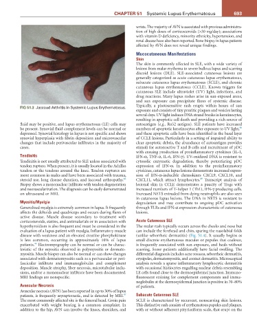
FIG 51.3 Jaccoud Arthritis in Systemic Lupus Erythematosus. Typically, a photosensitive rash erupts within hours of sun
exposure and consists of tiny pruritic plaques and vesicles lasting
several days. UV light induces DNA strand breaks in keratinocytes,
resulting in apoptotic cell death and providing a rich source of
fluid may be positive, and lupus erythematosus (LE) cells may autoantigen (e.g., Ro52 antigen). SLE patients have increased
48
be present. Synovial fluid complement levels can be normal or numbers of apoptotic keratinocytes after exposure to UV light,
depressed. Synovial histology in lupus is not specific and shows and these apoptotic cells have been identified in the basal layer
synovial hyperplasia with fibrin deposition and microvascular of CCLE lesions. Particularly in a setting of impaired ability to
changes that include perivascular infiltrates in the majority of clear apoptotic debris, the abundance of autoantigen provides
cases. stimuli for autoreactive T and B cells and recruitment of pDC
with ensuing production of proinflammatory cytokines (IL-1,
Tendinitis IFN-α, TNF-α, IL-6, IFN-γ). UV-oxidized DNA is resistant to
Tendinitis is not usually attributed to SLE unless associated with cytosolic enzymatic degradation, thereby potentiating pDC
tendon rupture. When present, it is usually located in the Achilles expression of IFN-α. In addition to the proinflammatory
tendon or the tendons around the knee. Tendon ruptures are cytokines, cutaneous lupus lesions demonstrate increased expres-
more common in males and have been associated with trauma, sion of IFN-α-inducible chemokines CXCL9, CXCL10, and
46
49
steroid use, long disease duration, and Jaccoud arthropathy. CXCL11, which attract lymphocytes. Transcript analysis of
Biopsy shows a mononuclear infiltrate with tendon degeneration lesional skin in CCLE demonstrates a paucity of Tregs with
and neovascularization. The diagnosis can be easily demonstrated increased numbers of T-helper 1 (Th1), IFN-γ producing cells.
on ultrasound or MRI. Increased NETS extruded from dying neutrophils are also seen
in cutaneous lupus lesions. The DNA in NETS is resistant to
Myositis/Myalgia degradation and may contribute to ongoing pDC activation
Generalized myalgia is extremely common in lupus. It frequently through TLRs and IFN-α expression characteristic of cutaneous
affects the deltoids and quadriceps and occurs during flares of lesions.
active disease. Muscle disease secondary to treatment with
corticosteroids, statins, and antimalarials or in association with Acute Cutaneous SLE
hypothyroidism is also frequent and must be considered in the The malar rash typically occurs across the cheeks and nose but
evaluation of a lupus patient with myalgia. Inflammatory muscle can include the forehead and chin, sparing the nasolabial folds
disease with weakness and an elevated creatine phosphokinase (unlike seborrheic dermatitis) (Fig. 51.4). It usually begins as
is less common, occurring in approximately 10% of lupus small discrete erythematous macules or papules that coalesce,
44
patients. Electromyography can be normal or can be charac- is frequently associated with sun exposure, and heals without
teristic of the myositis observed in polymyositis or dermato- scarring. Some patients additionally have facial swelling. The
myositis. Muscle biopsy can also be normal or can show changes differential diagnosis includes acne rosacea, seborrheic dermatitis,
associated with dermatomyositis such as a perivascular or peri- erysipelas, dermatomyositis, and contact dermatitis. Microscopical
fascicular infiltrate and immunoglobulin and complement analysis reveals a sparse inflammatory lymphocytic dermatitis
deposition. Muscle atrophy, fiber necrosis, microtubular inclu- with occasional histiocytes engulfing nuclear debris resembling
sions, and/or a mononuclear infiltrate have been documented. LE cells found close to the dermoepidermal junction. Immuno-
MRI findings are nonspecific. fluorescent staining for complement components and immu-
noglobulin at the dermoepidermal junction is positive in 70–80%
Avascular Necrosis of patients.
Avascular necrosis (AVN) has been reported in up to 30% of lupus
47
patients, is frequently asymptomatic, and is detected by MRI. Subacute Cutaneous SLE
The most commonly affected site is the femoral head. Groin pain SCLE is characterized by recurrent, nonscarring skin lesions.
exacerbated with weight bearing is a common complaint. In This distinctive rash consists of erythematous papules and plaques,
addition to the hip, AVN can involve the knees, shoulders, and with or without adherent pityriasiform scale, that erupt on the