
Page 739 - Clinical Immunology_ Principles and Practice ( PDFDrive )
P. 739
CHaPtEr 52 Rheumatoid Arthritis 711
proteins, fibrinogen fragments, antibody-DNA complexes, high- of inflammatory leukocytes, including polymorphonuclear
mobility group box (HMGB)-1, and hyaluronan oligosaccharides. leukocytes and immature or undifferentiated monocytes, orches-
Recent data suggest that synovial FLSs, as well as synovial trated by chemokines produced by resident stromal as well as
macrophages, express TLR2 in situ. Expression is upregulated infiltrating cells (Fig. 52.3). CXC, CC, C, and CX 3 C chemokines
after stimulation with IL-1 and the TLR2 ligand peptidoglycan. all play a role, exerting chemotactic activity toward neutrophils,
TLR2 engagement induces cytokines such as IL-6, matrix metal- lymphocytes, and monocytes but also influencing the topology
loproteinases, adhesion molecules, and an array of chemokines of inflammatory infiltrates. They are invariably early activation
including granulocyte chemotactic protein (GCP)-2, RANTES, genes (e.g., type I IFN response genes), in response to inflam-
monocyte chemoattractant protein (MCP)-2, IL-8, growth-related matory stimuli. Besides the homeostatic chemokines described
oncogene-2 and, to a lesser extent, macrophage-inflammatory above, the key players include IL-8/CXCL8, RANTES/CCL5,
protein 1α, MCP-1, EXODUS, and CXCL-16. Data suggest that MIP-1α/CCL3, SDF-1/CXCL12, IP-10/CXCL10, and MCP-1/
27
TLR3, TLR4, TLR7, and TLR9 are also expressed at messenger CCL2. Upregulation on endothelium of cell surface adhesion
25
ribonucleic acid (mRNA) and possibly protein level and may molecules, including ICAM-1, VCAM-1, and E-selectin, permits
augment inflammatory cytokine expression by DCs from patients the rolling and adhesion of leukocytes as they migrate. In synovial
with RA. joints, resident stromal cells and infiltrating macrophages are a
DCs are thought to be the most important antigen-presenting dominant source of such factors. Crucially, the expression of
cells in RA. Indeed, the proinflammatory environment favors cognate chemokine receptors such as CCR4, CCR5, CCR6, CXCR3,
DC maturation in regional lymph nodes as well as inflamed and CX3CR1 on inflammatory cell subsets contribute selectivity
26
27
tissue. Thus in peripheral blood, DC precursors express either of cellular recruitment. Within the T-cell compartment, there
−
dim
dim
an immature CD33 CD14 CD16 phenotype or a more mature exist distinct profiles of chemokine receptor expression, patterns
+
dim
MHC class II bright CD11c CD33 bright CD14 surface phenotype evolved perhaps to facilitate eradication of pathogens. For
typical of conventional myeloid DC (mDC); neither population example, T-helper 1 cells preferentially express CXCR3 and CCR5;
expresses costimulatory molecules. In contrast, synovial fluid Th2 cells express CCR3; Th17 cells express CCR6 and CCR4;
and tissue DC subsets resemble mature peripheral blood cells; and Tfh cells express CXCR5. Data suggest that CCR5, CCR6,
in addition, a subset expresses high levels of CD86 and can CCR7, CXCR3, CXCR4, and CXCR5 may all be important for
support allogeneic mixed leukocyte reactions. More recent data B-cell migration into synovium. Together, these events characterize
indicate that they may differentiate further in situ as suggested the acute phase of an innate immune response, a key checkpoint
by nuclear translocation of RelB in DC localized within peri- that precedes the progression to subsequent events that herald
vascular infiltrates, consistent with prior cytokine receptor or the onset of the chronic inflammatory phase.
TLR engagement in vivo. Perivascular RA synovium also contains
+
−
+
populations of MHC class II CD11c CD123 plasmacytoid DC Autoantigens in RA
(pDC); in contrast to the conventional myeloid DC subset, these Although current models of adaptive immune responses would
−
are RelB and comprise ~30% of all synovial DC. A subset suggest that DC carry antigens derived from damaged or dying
of pDC express BDCA2, capable of producing IFN-α in situ. synovial tissue, the molecular nature of disease-associated antigens
Unlike their peripheral blood counterparts, synovial pDC effi- has, until recently, remained an enigma. Many RA-associated
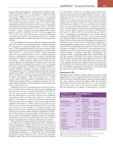
ciently activate allogeneic T cells to proliferate as well as to produce autoantigens have been described (see Table 52.1 for examples),
28
IFN-γ, TNF-α, and IL-10. and for some there exist in vivo arthritis model correlates.
Although the common myeloid precursor cell is the precursor The best described are collagen II, proteoglycans, HCgp-39,
for all myeloid cells, including DC and tissue macrophages, the
+
+
+
−
precise role of monocytes—namely CD14 CD16 , CD14 CD16 ,
+
dim
and the more recently described CD14 CD16 subset—in TABLE 52.1 autoantigens in
synovial inflammation is uncertain. They are good candidates rheumatoid arthritis
as persistence factors through their capacity to activate and
polarize T-cell subsets, to respond to the environment through t or B Molecular
TLR expression, and to produce a wide range of inflammatory Established Cell a Specificity assay b
mediators, including IL-1, TNF, IL-6, IL-8, CCL2, NO, and type Immunoglobulin G B Human Fc IgG Rheumatoid factor
+
dim
I IFN. In contrast a more recently described CD14 CD16 subset Cyclic peptides T and B Citrullinated Anti-CCPs
may function to sense damage, to scavenge cell debris and peptides
higher-order molecular complexes, and to promote angiogenesis Fibrin T and B α- and β-chain Research b
and tissue repair. More work is needed to establish the contribu- epitopes b
tion of these distinct functional subsets to the chronic inflam- Fibrinogen T and B Multiple epitopes Research b
T and B CEP-1 dominates Research
Enolase
matory process. Nonetheless, multiple cellular and molecular Vimentin T and B Citrullinated MCV assay
determinants exist in RA synovium that could serve both to vimentin
initiate and perpetuate the immune inflammatory response. Collagen II T and B Multiple epitopes Research b
This initial wave of inflammation has two major consequences. HnRNPA2 B Multiple epitopes Research b
First, inflammatory cytokines will promote the activation of Aggrecan T and B Multiple epitopes Research b b
vascular endothelium, changes that occur very early in disease HCgp-39 T Multiple epitopes Research b
Multiple epitopes Research
Glucose-6-phosphate B
(see above and Fig. 52.3). Under the influence of locally generated isomerase
cytokines and chemokines, synovial postcapillary venules undergo
morphological changes to an extent that they resemble high a b Denotes autoantigens recognized by T or B cells, or both.
Assay is either not commercially available or not in routine clinical use. Details of
endothelial venules similar to those observed in secondary assays may be found in primary research communications.
lymphoid organs. The second major consequence is the migration CCPs, cyclic citrullinated peptides; MCV, modified citrullinated vimentin.