
Page 756 - Clinical Immunology_ Principles and Practice ( PDFDrive )
P. 756
728 Part six Systemic Immune Diseases
lysozyme levels are useful when considering sarcoidosis as the
etiology of childhood arthritis and uveitis in early-onset sar-
coidosis cases, but noncaseating granulomas seen on biopsy are
much more revealing. As for imaging techniques, radiography
can be helpful in ruling out structural changes resulting from
disease processes other than arthritic disease. Nevertheless,
periarticular osteopenia is commonly seen around an inflamed
joint. The clinical presentation usually precedes the development
of bony erosions, but sclerosis of the sacroiliac joint may indicate
axial involvement in patients with ERA. The role of ultrasonog-
raphy for diagnosis and monitoring of disease progression in
pediatric patients is promising, but further establishment of
normative data is still required. MRI, with and without intra-
venous contrast, may help identify synovitis, but even this imaging
modality can sometimes yield false-positive results in certain
joints in otherwise healthy children. 8,21,22
DIFFERENTIAL DIAGNOSIS
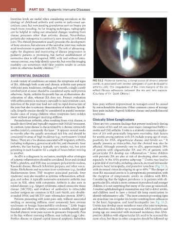
A wide variety of conditions can mimic the symptoms and signs FiG 53.2 Posterior synechia, a complication of chronic anterior
of JIA. Although both acute and chronic arthritis may present uveitis, is associated with several categories of juvenile idiopathic
with joint pain, tenderness, swelling, and warmth, a single acutely arthritis (JIA). The irregularities of the inner margins of the iris
involved joint at onset should be considered septic until proven reflect fibrous adhesions between the iris and lens capsule.
otherwise. Septic arthritis frequently has an erythematous dis- (Courtesy of Dr. Scott Olitsky.)
coloration of skin, whereas JIA does not. Prompt evaluation
with arthrocentesis is necessary, especially in indeterminate cases.
Infection of the joint may lead not only to rapid destruction of knee pain without improvement in teenagers could be caused
the joint but also to systemic dissemination of infection. Specifi- by osteochondritis dissecans. Other common causes of teenage
cally, Kingella species should be considered in painful monoar- knee pain include Osgood-Schlatter disease and patellofemoral
ticular cases of young children when symptoms have sudden syndrome.
onset without prolonged morning stiffness.
Parainfectious arthritis, often resulting from viral disease, is Clinically Silent Complications
usually short-lived and typically requires only NSAID treatment. There are two common findings that present insidiously during
In contrast, Lyme disease is characterized by a chronic extensively the course of JIA and yet can cause major damage/morbidity—
13
swollen joint(s), commonly the knee. It appears several weeks uveitis and TMJ arthritis. Uveitis is a relatively common complica-
to months after the usually unnoticed tick bite and should be tion of JIA with potentially long-term morbidity. Risk factors
considered in areas of high incidence (e.g., northeastern United for uveitis among patients with JIA include young age of onset,
14
States). There are a few diseases associated with migratory arthritis, positivity for ANA, oligoarticular disease, and female sex. It
including malignancy, gonococcal arthritis, and rheumatic fever usually presents as iridocyclitis, but the choroid may also be
arthritis, the last having a typically very tender, red, hot joint affected. Although extremely rare in sJIA, approximately 20%
persisting in each location for a couple of hours before moving of patients with oligoarticular JIA and 5% of patients with
14
to another. polyarticular JIA develop eye inflammation. Some children
As sJIA is a diagnosis by exclusion, multiple other etiologies with psoriatic JIA are also at risk of developing silent uveitis,
19
of systemic inflammation should be considered. Fever and elevated especially in the ANA-positive subgroup. Uveitis may lead to
WBCs, platelets, and ESR may accompany polyarteritis nodosa, a great deal of morbidity, including cataracts, increased intraocular
Kawasaki disease, Henoch-Schönlein purpura, and other vascu- pressure, band keratopathy, and posterior synechiae (Fig. 53.2),
14
litides. Ehrlichiosis and recurrent fever syndromes (e.g., familial with decreased vision developing in up to 40%. The danger of
Mediterranean fever, TNF receptor–associated periodic fever most JIA-associated uveitis is its asymptomatic presentation, with
syndrome) may also manifest as systemic inflammation, arthral- the exception of symptomatic uveitis in children with ERA.
gias, and rashes. A typically nonerosive symmetric arthritis may Considering that the highest prevalence is in patients with oli-
occur in SLE, often with cytopenias. Arthritis is also seen in goarticular JIA, which is most common in preschool and younger
related diseases (e.g., Sjögren syndrome, mixed connective tissue children, it is not surprising that many of the cases go unnoticed.
disease [MCTD]), and evidence of antibodies to extractable A routine ophthalmological examination may fail to detect uveitis,
nuclear antigens are common in SLE (anti-Smith, anti–double- and children need to have a formal slit-lamp examination to
14
stranded DNA) and MCTD (anti–ribonuclear protein [RNP]). identify inflammatory cells. The most common presenting signs
Patients presenting with joint pain only, without associated are synechiae (an irregular iris border resulting from adhesions
swelling or morning stiffness, most commonly have overuse to the lens), hypopyon, and band keratopathy (see Fig. 53.2).
23
syndromes or benign hypermobility syndrome. Little league Uveitis may develop many months or years after joint symptoms,
shoulder and elbow are rather common in middle school–aged and therefore close follow-up is warranted. ANA positivity and
baseball players; wrist pain occurs in gymnasts frequently. Pain young age are associated with increased incidence; thus ANA-
in the hip, without morning stiffness, may indicate Legg-Calve- positive children with oligoarticular JIA need to be screened the
Perthes disease or slipped capital femoral epiphysis. Relentless most often, but those in other categories should be followed up