
Page 868 - Clinical Immunology_ Principles and Practice ( PDFDrive )
P. 868
838 ParT Six Systemic Immune Diseases
TABLE 61.5 Preliminary Criteria for the
Classification of Catastrophic
antiphospholipid Syndrome
1. Evidence of involvement of three or more organs, systems, and/or
tissues a
2. Development of manifestations simultaneously or in less than 1
week
3. Confirmation by histopathology of small-vessel occlusion in at least
one organ or tissue b
4. Laboratory confirmation of the presence of antiphospholipid
antibody (aPL) c
Definite Catastrophic antiphospholipid Syndrome (aPS):
• All 4 criteria
Probable Catastrophic aPS:
• Criteria 2–4 and two organs, systems, and/or tissues involved;
• Criteria 1–3, except no aPL confirmation 6 weeks apart due to the
early death of a patient not tested before catastrophic episode;
• Criteria 1, 2, 4; or
• Criteria 1, 3, 4, and development of a third event more than 1
week but less than 1 month after first despite anticoagulation.
a Usually, clinical evidence of vessel occlusions, confirmed by imaging techniques
when appropriate. Renal involvement is defined by a 50% rise in serum creatinine,
severe systemic hypertension, and/or proteinuria.
b For histopathological confirmation, significant evidence of thrombosis must be
present, although vasculitis may coexist occasionally.
c If the patient had not been previously diagnosed as having APS, laboratory
confirmation requires that the presence of aPL must be detected on two or more
occasions at least 12 weeks apart (not necessarily at the time of the event),
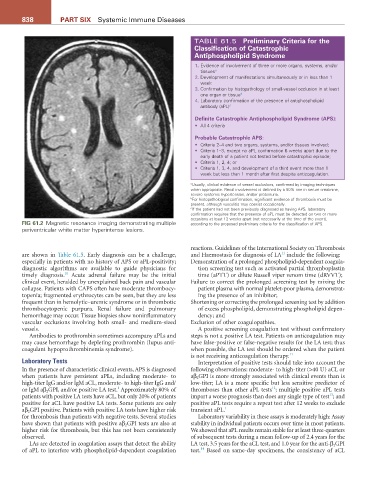
FiG 61.2 Magnetic resonance imaging demonstrating multiple according to the proposed preliminary criteria for the classification of APS.
periventricular white matter hyperintense lesions.
reactions. Guidelines of the International Society on Thrombosis
11
are shown in Table 61.5. Early diagnosis can be a challenge, and Haemostasis for diagnosis of LA include the following:
especially in patients with no history of APS or aPL-positivity; Demonstration of a prolonged phospholipid-dependent coagula-
diagnostic algorithms are available to guide physicians for tion screening test such as activated partial thromboplastin
10
timely diagnosis. Acute adrenal failure may be the initial time (aPTT) or dilute Russell viper venom time (dRVVT);
clinical event, heralded by unexplained back pain and vascular Failure to correct the prolonged screening test by mixing the
collapse. Patients with CAPS often have moderate thrombocy- patient plasma with normal platelet-poor plasma, demonstrat-
topenia; fragmented erythrocytes can be seen, but they are less ing the presence of an inhibitor;
frequent than in hemolytic–uremic syndrome or in thrombotic Shortening or correcting the prolonged screening test by addition
thrombocytopenic purpura. Renal failure and pulmonary of excess phospholipid, demonstrating phospholipid depen-
hemorrhage may occur. Tissue biopsies show noninflammatory dency; and
vascular occlusions involving both small- and medium-sized Exclusion of other coagulopathies.
vessels. A positive screening coagulation test without confirmatory
Antibodies to prothrombin sometimes accompany aPLs and steps is not a positive LA test. Patients on anticoagulation may
may cause hemorrhage by depleting prothrombin (lupus anti- have false-positive or false-negative results for the LA test; thus
coagulant hypoprothrombinemia syndrome). when possible, the LA test should be ordered when the patient
is not receiving anticoagulation therapy. 11
Laboratory Tests Interpretation of positive tests should take into account the
In the presence of characteristic clinical events, APS is diagnosed following observations: moderate- to high-titer (>40 U) aCL or
when patients have persistent aPLs, including moderate- to aβ 2 GPI is more strongly associated with clinical events than is
high-titer IgG and/or IgM aCL, moderate- to high-titer IgG and/ low-titer; LA is a more specific but less sensitive predictor of
12
1
or IgM aβ 2 GPI, and/or positive LA test. Approximately 80% of thromboses than other aPL tests ; multiple positive aPL tests
13
patients with positive LA tests have aCL, but only 20% of patients impart a worse prognosis than does any single type of test ; and
positive for aCL have positive LA tests. Some patients are only positive aPL tests require a repeat test after 12 weeks to exclude
aβ 2 GPI positive. Patients with positive LA tests have higher risk transient aPL. 1
for thrombosis than patients with negative tests. Several studies Laboratory variability in these assays is moderately high: Assay
have shown that patients with positive aβ 2 GPI tests are also at stability in individual patients occurs over time in most patients.
higher risk for thrombosis, but this has not been consistently We showed that aPL results remain stable for at least three-quarters
observed. of subsequent tests during a mean follow-up of 2.4 years for the
LAs are detected in coagulation assays that detect the ability LA test, 3.5 years for the aCL test, and 1.0 year for the anti-β 2 GPI
14
of aPL to interfere with phospholipid-dependent coagulation test. Based on same-day specimens, the consistency of aCL