
Page 890 - Clinical Immunology_ Principles and Practice ( PDFDrive )
P. 890
CHaPtEr 63 Bullous Diseases of the Skin and Mucous Membranes 861
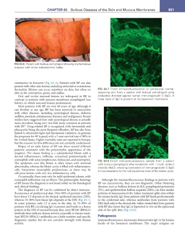
FIG 63.6 Patient with bullous pemphigoid showing erythematous
plaques with tense subepidermal bullae.
centimeters in diameter (Fig. 63. 6). Patients with BP can also
present with other skin lesions, including urticarial or eczematous
dermatitis. Blisters can occur anywhere on skin, but often on FIG 63.7 Direct immunofluorescence of perilesional normal-
skin in the extremities, groin, and axillae. appearing skin from a patient with bullous pemphigoid using
Oral and ocular mucosal lesions are infrequent in BP, in antibodies directed against human immunoglobulin G (IgG). A
contrast to patients with mucous membrane pemphigoid (see linear band of IgG is present at the basement membrane.
below), in which mucosal lesions predominate.
Most patients with BP are over 60 years of age, although it
can develop at any age. BP has been reported in association
with other diseases, including neurological disease, diabetes
mellitus, psoriasis, autoimmune diseases, and malignancy. Recent
studies have suggested that only neurological disease is actually
more prevalent, being over two-fold more common in patients
25
with BP. Drug-related BP is recognized, with furosemide and
phenacetin being the most frequent offenders. BP has also been
linked to ultraviolet light and therapeutic radiation. In general,
the prognosis for BP is good, with a 1-year survival rate of 90% in
the United States. Higher mortality rates are reported in Europe,
but the reasons for this difference are not currently understood.
Biopsy of an early lesion of BP can show several different
patterns consistent with the polymorphic appearance of the
eruption. The classic finding is a subepidermal blister with a
dermal inflammatory infiltrate, comprised predominantly of
eosinophils with some lymphocytes, histiocytes, and neutrophils. FIG 63.8 Direct immunofluorescence sample from a patient
The epidermis over this blister is often intact with minimal with bullous pemphigoid after incubation with 1 mol/L sodium
abnormality, whereas the blister cavity is filled with inflammatory chloride (NaCl), showing localization of immunoglobulin G (IgG)
cells. Sometimes neutrophils predominate, or there may be immunoreactants to the roof (epidermal side) of the blister cavity.
cell-poor lesions with very few inflammatory cells.
Occasionally, there may only be mild epidermal edema, with
eosinophil infiltration but no blister. The polymorphic histology Although the immunofluorescence findings in patients with
of BP means the diagnosis is not based solely on the histological BP are characteristic, they are not diagnostic. Other blistering
and clinical findings. diseases, such as bullous lesions in SLE, pemphigoid gestationis
The diagnosis of BP can be confirmed by direct immuno- (PG), and epidermolysis bullosa acquisita (EBA), can have similar
fluorescence of perilesional skin. Over 90% of patients with BP patterns of immunoreactivity. Saline treatment splits skin within
have linear C3 deposits at the dermal–epidermal junction (DEJ), the lamina lucida: IgG from patients with BP binds predominantly
whereas 70–90% have linear IgG deposits at the DEJ (Fig. 63.7). to the epidermal side, whereas antibodies from patients with
In some patients, only C3 is seen in the skin. In 70–90% of EBA bind only to the dermal side. Saline-treated skin from patients
patients with BP, circulating IgG is present and binds in a linear with BP also shows that IgG is deposited in vivo on the epidermal
pattern at the DEJ of normal human skin. Anti-BP180 (BPAG2) side of the split skin (Fig. 63.8).
antibody titers indicate disease activity, especially at disease onset.
Anti-BP230 (BPAG1) antibodies are a fairly sensitive and specific Pathogenesis
diagnostic marker but do not correlate strongly with disease Immunofluorescence microscopy demonstrates IgG in the lamina
activity. lucida of the basement membrane. The target antigens are