
Page 892 - Clinical Immunology_ Principles and Practice ( PDFDrive )
P. 892
CHaPtEr 63 Bullous Diseases of the Skin and Mucous Membranes 863
EPIDERMOLYSIS BULLOSA ACQUISITA in BP. Linear deposits of C3, IgM, IgA, and fibrinogen have also
35
been reported. However, in EBA, these deposits are localized
EBA is a chronic subepidermal blistering disease that typically exclusively below the lamina lucida. Direct immunofluorescence
presents in the fourth to sixth decades. There are two forms of of saline-split perilesional skin shows IgG in the blister floor in
EBA—a noninflammatory type with blisters on distal extremities EBA (Fig. 63.10); in BP, IgG is in the blister roof.
and an inflammatory type that closely resembles BP. Patients Indirect immunofluorescence using normal human skin shows
with non-inflammatory EBA have peripherally distributed blisters circulating anti–basement membrane zone antibodies in 30–50%
that heal with scarring and milia formation. Their skin is extremely of patients with EBA. However, IgG antibody can be detected
fragile, often resulting in numerous erosions in areas of mechanical in 85% of patients when using saline-split skin, which is the
trauma, such as hands, feet, elbows, and knees. Lesions are often more sensitive and specific substrate. As expected from the in
seen on oral mucous membranes, sometimes including the vivo deposition pattern, IgG from patients with EBA localizes
esophagus. Patients with classic EBA may also develop ocular, to the blister floor. Sera from these patients recognize the 300-kDa
vaginal, urethral, and rectal mucosal lesions. Ocular changes are protein, type VII collagen, primarily targeting its immunodomi-
common, clinically resembling mucous membrane pemphigoid nant NC1 domain.
(MMP). Other cutaneous manifestations include scarring alopecia
and variable degrees of nail dystrophy. Patients with nonclassic Pathogenesis
(inflammatory) EBA often present similarly to those with BP, Immunoelectron microscopy of EBA biopsy specimens has shown
with widespread tense bullae on an erythematous base, which immunoglobulin deposits localized to the lamina densa zone of
heal without scarring (Fig. 63.9). 35 the basement membrane, below the lamina lucida. Transmission
EBA has been associated with several diseases, particularly electron microscopy of lesional EBA skin shows decreased or
inflammatory bowel disease (IBD) and bullous SLE. These absent anchoring fibrils. Anchoring fibrils are implicated in
associations may partly result from the association of EBA with epidermal–dermal adherence via linkage of the hemidesmosome
HLA-DR2. 36 through the basement membrane, and their absence may explain
Skin biopsies of early lesions from patients with EBA show the observed skin fragility. The lack of inflammatory infiltrate
subepidermal blisters with variable degrees of inflammation. In in many patients with EBA suggests that autoantibodies may
patients with classic EBA, lesional skin biopsies often have minimal disrupt the interaction between anchoring fibrils and dermal
inflammatory cell infiltrate. In contrast, patients with inflam- matrix proteins.
matory EBA may have substantial collections of mononuclear The target antigen for the IgG autoantibodies present in the
cells, neutrophils, and eosinophils in the superficial dermis. sera of patients with EBA is type VII collagen, a 300-kDa gly-
Direct immunofluorescence of perilesional skin biopsies from coprotein composed of a 145-kDa noncollagenous domain (NC1)
patients with EBA shows linear deposits of IgG at the DEJ, as at the amino-terminal end, an 18-kDa noncollagenous domain
(NC2) at the carboxy terminus, and a central collagenous domain.
IgG antibodies from patients with EBA appear specific for epitopes
within the NC1 noncollagenous domain. Type VII collagen is
the major structural component of anchoring fibrils and is
produced by both epithelial keratinocytes and dermal fibroblasts.
Passive transfer experiments in mice and active immune models
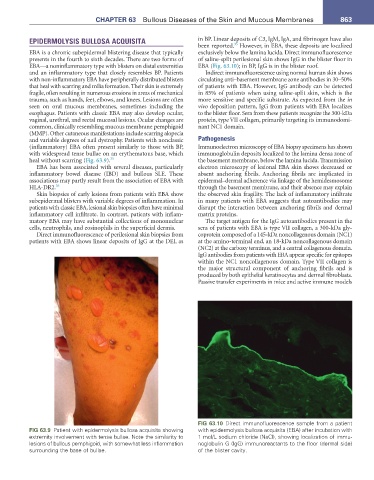
FIG 63.10 Direct immunofluorescence sample from a patient
FIG 63.9 Patient with epidermolysis bullosa acquisita showing with epidermolysis bullosa acquisita (EBA) after incubation with
extremity involvement with tense bullae. Note the similarity to 1 mol/L sodium chloride (NaCl), showing localization of immu-
lesions of bullous pemphigoid, with somewhat less inflammation noglobulin G (IgG) immunoreactants to the floor (dermal side)
surrounding the base of bullae. of the blister cavity.