
Page 1162 - Hall et al (2015) Principles of Critical Care-McGraw-Hill
P. 1162
CHAPTER 86: Intracranial Pressure: Monitoring and Management 801
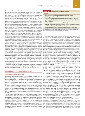
the above findings as ICP is either normalized or worsens. As a result, TABLE 86-7 Conditions Often Requiring ICP Monitoring
frequent reexamination is necessary to assess the patient’s response to
therapeutic measures. Increasing downward pressure leads to dysfunc- Conditions
tion first at the level of the diencephalon (ie, thalami), next affecting • Comatose patient, unexplained and/or with abnormal imaging findings
the upper and middle sections of the brainstem (midbrain and pons), • Clinical symptoms of elevated ICP
and ultimately impeding medullary function. In summary of the above • In TBI, normal CT scan with more than two of the following features noted at admission:
examination methods, assessment of clinical signs tracks the descend- age >40 y/o, unilateral or bilateral motor posturing, or systolic blood pressure <90 mm Hg.
ing progression of injury in evolving intracranial hypertension. The first • Diffuse brain edema
is assessment of the respiratory pattern. Respiration patterns become • Extensive hemispheric brain edema (eg, large MCA infarct)
progressively more abnormal depending on the level of injury, evolving • Intracerebral hemorrhage with edema causing significant mass effect (clinical and radiographic)
from a Cheyne-Stokes pattern to ataxic respirations and then eventu- • Contusion and edema especially with bifrontal and temporal involvement
ally to apnea. The pupils become increasingly more abnormal, initially • Radiographic evidence of ventriculomegaly with clinical evidence of hydrocephalus
with some early constriction, then increasing dilation and diminished
reactivity, to ultimately becoming fixed, unreactive, and middilated CT, cranial tomography; ICP, intracranial pressure; MCA, middle cerebral artery; TBI, traumatic brain
injury. Common conditions that may necessitate direct ICP monitoring.
(Fig. 86-14). Reflex eye movements (ie, doll’s eye maneuver) are even-
tually lost (pontine compromise) (Fig. 86-14 and Table 86-5). Motor
responses evolve from localizing to nonlocalizing withdrawal followed
by decorticate and then decerebrate posturing and finally flaccidity of Regarding appropriate selection of patients for invasive ICP
all extremities (Table 86-5). The end result of untreated, progressive monitoring, the best ICP guidelines are found in the latest Brain Trauma
rostrocaudal brain herniation is brain death with loss of all cranial nerve Foundation recommendations (level II evidence). These guidelines
reflexes and no respiration in the setting of elevated CO . 2 indicate the use of ICP monitoring in patients with TBI who remain
Depending on the level of coma or sedation, the patient may not comatose after resuscitation and if the admission CT reveals intracranial
react to any stimulus, or may respond to noxious or painful stimuli by pathology such as hematoma, contusions, or brain edema. Generally
32
grimacing, grabbing, or withdrawing the stimulated body part. Patients accepted indications for monitor insertion are processes associated
should be stimulated in all extremities to compare the response on both with progressive elevation of ICP such as rapidly expanding intracranial
sides in order to attempt to localize the etiology of brain dysfunction. If masses secondary to ischemia, hematoma, hemorrhagic tumor, obstruc-
a patient’s response is equivalent throughout all extremities, it is likely tive and nonobstructive hydrocephalus, or diffuse axonal injury (DAI).
that the etiology is a global insult. If there is a focal response, this finding In all of these forms of brain injury, treatment demands active pres-
can help refine the anatomic location of the injury as discussed above. ervation of stable CPP and ICP to maintain adequate brain perfusion.
Sensory stimuli may be delivered by pinching a small skin area at the Neuroimaging studies assist in determining the indication for ICP
mediolateral forearm or inner thigh; sometimes, however, nail bed pres- monitoring. For example, identification of midline shift, effacement of
sure is needed. These maneuvers should be avoided in patients with a the basal cisterns, or extensive edema helps narrow the differential diag-
coagulopathy; as an alternative, pressure applied over the supraorbital nosis to a process involving elevated ICP. Significant ICP elevations may
notch, bilateral mastoids, or cervicospinal muscles can be used. Most occur without, or with only subtle, brain imaging findings. Therefore,
elegantly, some patients respond strongly and reproducibly to intranasal imaging studies should always be interpreted together with the clinical
stimulations with a cotton swab. findings and brain monitoring information. ICP monitoring indications
Frequent bedside examination by physicians and nurses looking for are listed in Table 86-7.
these abnormal findings should be performed in all patients at risk for To understand the potential benefit of ICP-based treatment algorithms
ICP elevations and the results tracked hourly in the patient’s chart. in TBI, Chesnut and colleagues prospectively studied 324 patients
33
in Bolivia and Ecuador using random assignment to manage severe
TBI patients (GCS 3-8), based on either serial CT imaging and clinical
INTRACRANIAL PRESSURE MONITORING examination (ICE) only or ICE plus invasive ICP monitoring (keeping
■ INDICATIONS FOR ICP MONITORING ICP <20 mm Hg). There was no significant difference in the primary
outcome, a composite measure based on percentile performance across
ICP monitoring can be an extremely important tool in managing patients 21 measures of functional and cognitive status. Mortality at 6 months
in acute brain injury and suspected intracranial hypertension as it was similar—41% versus 39% (p = 0.60), as was median length of ICU
provides information on a minute-to-minute basis. The challenges stay and distribution of serious adverse events. However, the number of
associated with invasive monitoring are the selection of appropriate brain-specific ICU treatment days (eg, use of hyperosmolar fluids and
patients, and the accurate analysis of the information provided by the hyperventilation) was lower in the ICE plus ICP than the ICE group
probe. As with all invasive monitors, the waveform and values provided (3.4 vs 4.8 days; p = 0.002). Taken together, these results seem to support
by an ICP probe should be carefully interpreted, as inaccurate analysis the lack of superiority of an ICP treatment algorithm over treatment
of pressure waveforms can be potentially dangerous. Of note, both the solely guided by ICE only in severe TBI patients. The authors concluded
value of the ICP and its waveform provide important information and that although there were no outcome differences, the qualitative (not
furthermore, the waveform can indicate worsening pressure dynamics quantitative) nature of the ICE-only approach and the increased treat-
via the Lundberg waves, information that may not be communicated by ment efficiency (ie, tailoring osmotherapy) in the ICE plus ICP group
the ICP value alone. should not change the practice of ICP monitoring in areas where this
Invasive ICP monitoring should be used in the setting of a clini- resource is available. Further, the authors mentioned that their findings
cal examination that raises concern for intracranial hypertension or do not argue against the use of ICP monitoring as only the monitoring-
a mechanism of brain injury or radiographic findings that would be based interventional algorithm was tested in their study.
consistent with elevated ICP, especially in a patient who is unable to be Four important discussion points should be considered prior to read-
appropriately examined due to sedation or paralysis. In such patients, ing the study results and the impact on current and modern ICP man-
“blind” management without an ICP monitor can actually exacerbate agement strategies: the study location and scope of practice in Bolivia
37
secondary injury and result in a worse outcome for the patient. For and Ecuador; physicians’ expertise and complication rates; variations in
example, uncontrolled use of hyperventilation and osmotic therapy ICP interpretation and management skills; variations among the severe
without ICP guidance may lead to decreased CBF and resultant regional TBI patients; and, lastly, the monitoring device employed and the fact
or global ischemia. that other reasonable indications for ICP monitoring exist. First, it is
section06.indd 801 1/23/2015 12:56:02 PM