
Page 1477 - Hall et al (2015) Principles of Critical Care-McGraw-Hill
P. 1477
1016 PART 9: Gastrointestinal Disorders
A rebleeding. Studies comparing standard-dose PPIs given by intermittent
intravenous infusion once or twice daily to continuous high-dose infu-
Pylorus
sion over 72 hours have shown equal efficacy of both regimens, with
similar rebleeding rates and mortality. 67,68 This suggests that intermittent
Gastric dosing may be an equally efficacious regimen for prevention of bleeding.
antrum Splanchnic vasoconstrictors such as somatostatin and octreotide are not
routinely recommended for patients with acute ulcer bleeding. These
16
can be considered in patients that cannot get endoscopy for any reason,
or for those with profuse bleeding awaiting surgery.
Angiography: In most cases of nonvariceal hemorrhage, endoscopic
evaluation is able to visualize the bleeding lesion and deliver effective
hemostatic therapy. However, in a minority of patients, the bleeding
source is not visualized by endoscopy, thereby necessitating angio-
graphic localization. Also, angiography offers the option of hemostatic
therapy using arterial vasoconstrictors or embolization; however, this
generally is reserved for patients who are poor surgical candidates or for
the control of bleeding in an unstable patient awaiting surgery.
Bleeding ulcer Localization As mentioned previously, angiography can successfully local-
ize brisk UGI bleeding (rate >0.5 mL/min) in 75% of cases, with
69
most bleeding episodes (85%) originating from a branch of the left
B gastric artery. The right gastric and short gastric arteries account for the
remainder of the sources.
Pylorus
Hemostatic Therapy UGI arterial bleeding can be controlled by the selective
arterial infusion of vasoconstrictors such as vasopressin or embolization
of particulate matter. Most studies indicate that selective intra-arterial
vasopressin is more effective than a peripheral intravenous infusion in
achieving hemostasis. Since the vasoconstrictive action of vasopres-
70
sin is more pronounced on terminal blood vessels such as arterioles,
venules, and capillaries, this therapy is more effective in controlling
bleeding from such vessels, as in diffuse hemorrhagic gastritis, rather
than a duodenal ulcer bleed originating from a large gastroduodenal
artery. In the setting of gastric bleeding, therapy appears to be equally
71
Hemostasis at effective when administered via the left gastric or celiac artery. A usual
bleeding site therapeutic vasopressin dose is 0.2 unit/min, with a recommended
maximum dose of 0.4 unit/min. If hemostasis is achieved, the infu-
sion is continued in the ICU for 24 to 36 hours and then tapered over
24 hours. Rebleeding after cessation of the infusion is a concern and
71
can be treated with repeat vasopressin treatment or embolization.
Embolization therapy uses various substances, most frequently a gela-
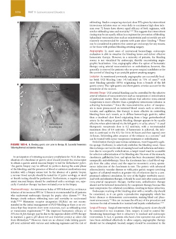
FIGURE 105-4. A. Bleeding gastric ulcer prior to therapy. B. Successful hemostasis tin sponge (Gelfoam), to selectively embolize the bleeding vessel. Since
following thermal and injection therapy. this technique carries the risk of causing bowel wall ischemia and infarc-
tion due to nonspecific embolization, a target vessel must be accessible
for selective catheterization of the bleeding site. Necrosis of the stomach,
In anticipation of initiating secondary prophylaxis for PUD, the visu- duodenum, gallbladder, liver, and spleen has been documented following
alization of a duodenal or gastric ulcer should prompt the endoscopist nonspecific embolotherapy. Since the duodenum has a dual blood sup-
to obtain a gastric antral mucosal biopsy to test for Helicobacter pylori. ply from the celiac artery and the superior mesenteric artery, sponta-
The gastric biopsy may be difficult to perform during the acute phase neous infarction of the duodenum is rare. The patient with advanced
of the bleeding, and the presence of blood or antiulcer medications may atherosclerotic vascular disease or with prior gastric surgery involving
interfere with a biopsy urease test. In the absence of a gastric biopsy, ligation of collateral vessels is at greater risk of infarction due to a com-
a venous blood sample should be tested for H pylori serology, or stool promised collateral circulation. In view of the higher morbidity associ-
or breath testing should be performed. Furthermore, a negative gastric ated with embolization therapy, it should be used only after unsuccessful
antral mucosal biopsy should be confirmed with a serologic test, espe- intra-arterial vasopressin therapy. Furthermore, embolization therapy
cially if antiulcer therapy has been initiated prior to the biopsy. should not be followed immediately by vasopressin therapy because this
Pharmacotherapy: An intravenous bolus of PPI followed by continuous may compromise the collateral circulation, resulting in tissue infarction.
infusion of intravenous PPI for 72 hours is recommended in all patients Endoscopic marking of the bleeding ulcer with a metallic clip should
who underwent endoscopic therapy for peptic ulcer disease. 16,63 This be considered, as it can guide superselective angiography which has
therapy clearly reduces rebleeding rates and mortality in randomized better chances to demonstrate extravasation, making blind coil place-
72
trials. 22,58,64 Histamine receptor antagonists (H RAs) are not recom- ment unnecessary. This can increase the efficacy of the procedure and
2
73,74
mended in the initial management of PUD bleeding as there is no evi- decrease the risk of nonselective luminal and hepatic embolization.
dence that they improve short-term outcomes, such as rebleeding rates Surgical Therapy: Surgical intervention for bleeding peptic ulcers should
or transfusion requirements. 57,65 The improved hemostatic efficacy of be considered in two situations. First, surgery is used to control life-
PPI over H RA therapy may be due to the superior ability of PPI therapy threatening hemorrhage that is refractory to medical and endoscopic
2
to maintain a gastric pH above 6.0 and therefore protect an ulcer clot intervention. In fact, in patients who have a low operative risk and who
from fibrinolysis. However, there are no clinical trials linking gastric have been stabilized effectively to allow surgery, angiographic therapy
66
pH level achieved with various acid reducing regimens and the risk of should not be attempted. Second, surgery should be considered in the
section09.indd 1016 1/14/2015 9:27:13 AM