
Page 1563 - Hall et al (2015) Principles of Critical Care-McGraw-Hill
P. 1563
1082 PART 10: The Surgical Patient
or other pliable material to the fascia or skin to prevent postoperative frequently have multiple intra-abdominal fluid collections and free
evisceration, and the wound is packed with saline-soaked gauze. 28-30 air that could be a result of the laparotomy. If the patient displays any
These patients are particularly at risk for the formation of enterocuta- signs of sepsis, such as fever or leukocytosis, despite being treated with
neous fistulae at the surface of their open wounds, and this complica- antibiotics, another search for the fever is warranted. However, the
31
tion is easily diagnosed by inspection. Tubes should be inspected to intensivist must keep in mind there are many noninfectious causes for
28
make sure that they have not been dislodged and are functioning as fever and leukocytosis in the ICU patient. The patient who is dete-
31
intended. For example, sump drains should be checked to make sure riorating in the first week following surgery for peritonitis and who
that the air inlet ports are not occluded. is thought to have persistent or recurrent peritoneal infection usually
Second, the intensivist must determine whether the gastrointestinal requires repeat laparotomy for source control. Beyond this early phase,
tract is functioning well enough for enteral feedings, difficult to deter- when abscesses are better formed and sterile collections have been
mine in the sedated, ventilated patient, and it is frequently necessary resorbed, image-guided percutaneous drainage offers a safe and effec-
https://kat.cr/user/tahir99/
to challenge the patient by starting tube feedings and simply checking tive method to diagnose and control abscesses.
the gastric residual volume every 4 hours. As discussed in Chap. 20, Open Abdomen Treatment In certain circumstances, patients with peri-
enteral feeding is preferred over parenteral feeding in this patient tonitis require “open abdomen” treatment. The skin and fascia are
population, if technically feasible. Jejunal feeds are almost always tol- not closed and evisceration is prevented by suturing an artificial
erated, even in patients with severe peritonitis. Early enteral feeding mesh or other flexible soft material to the fascia or skin. 28-30 These
may improve outcome. Enteral nutrition has not improved survival patients fall into two categories—patients whose fascia could not
25
but has reduced infectious morbidity—specifically intra-abdominal be closed for technical reasons but who are otherwise stable and
abscess in trauma patients. Before starting enteral feeds, it is neces- patients whose peritonitis is so severe that in the surgeon’s opinion
25
sary to ensure the bowel is in continuity. Occasionally surgeons may the abdomen should be left open to facilitate repeated laparotomies
first perform a “damage control” operation and leave the bowel in dis- for peritoneal toilet. The latter group presents a major problem to the
continuity with the intent of doing another laparotomy for definitive intensivist and the ICU nursing staff. These patients may undergo
repair in 24 to 72 hours. relaparotomy (through the mesh) every 1 to 3 days until the surgeon
Third, the intensivist must determine if the patient is septic and feels that the peritoneal cavity is sufficiently clean. Weaning from
if the septic focus is intra-abdominal. The most common abdomi- ventilatory support is almost always impossible until after the last
nal complication of peritonitis surgery is abscess formation, which scheduled relaparotomy. Furthermore, during this period of repeated
occurred in 21 of the 107 peritonitis patients who required postopera- laparotomies, large quantities of proteinaceous fluids are lost through
tive ventilation in our series. It is often difficult to determine when the open abdominal wound, and the patients may therefore require
21
a patient’s original septic response is abating and when a new septic support with aggressive nutrition. 25
response is being mounted. In general, if the patient is not improving
way, a CT scan of the abdomen (with IV contrast, if possible) should ■ VISCERAL ABSCESS
steadily following surgery or if the patient begins to deteriorate in any
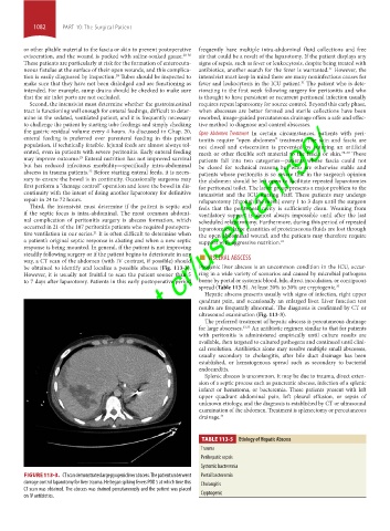
be obtained to identify and localize a possible abscess (Fig. 113-3). Pyogenic liver abscess is an uncommon condition in the ICU, occur-
However, it is usually not fruitful to scan the patient sooner than 5 ring in a wide variety of scenarios and caused by microbial pathogens
to 7 days after laparotomy. Patients in this early postoperative period borne by portal or systemic blood, bile, direct inoculation, or contiguous
spread (Table 113-5). At least 20% to 30% are cryptogenic. 32
Hepatic abscess presents usually with signs of infection, right upper
quadrant pain, and occasionally an enlarged liver. Liver function test
results are frequently abnormal. The diagnosis is confirmed by CT or
ultrasound examination (Fig. 113-3).
The preferred treatment of hepatic abscess is percutaneous drainage
for large abscesses. 32,33 An antibiotic regimen similar to that for patients
with peritonitis is administered empirically until culture results are
available, then targeted to cultured pathogens and continued until clini-
cal resolution. Antibiotics alone may resolve multiple small abscesses,
usually secondary to cholangitis, after bile duct drainage has been
established, or hematogenous spread such as secondary to bacterial
endocarditis.
Splenic abscess is uncommon. It may be due to trauma, direct exten-
sion of a septic process such as pancreatic abscess, infection of a splenic
infarct or hematoma, or bacteremia. These patients present with left
upper quadrant abdominal pain, left pleural effusion, or sepsis of
unknown etiology, and the diagnosis is established by CT or ultrasound
examination of the abdomen. Treatment is splenectomy or percutaneous
drainage. 34
TABLE 113-5 Etiology of Hepatic Abscess
Trauma
Perihepatic sepsis
Systemic bacteremia
FIGURE 113-3. CT scan demonstrates large pyogenic liver abscess. The patient underwent Portal bacteremia
damage control laparotomy for liver trauma. He began spiking fevers POD 5 at which time this Cholangitis
CT scan was obtained. The abscess was drained percutaneously and the patient was placed
on IV antibiotics. Cryptogenic
section10.indd 1082 1/20/2015 9:19:49 AM