Page 453 - Clinical Hematology_ Theory _ Procedures ( PDFDrive )
P. 453

CHAPTER 22  ■  Lymphoid and Plasma Cell Neoplasms                                  437


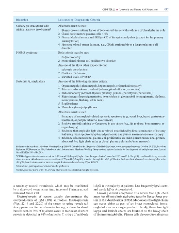
                       Disorder                                                Laboratory Diagnostic Criteria


                       Solitary plasmacytoma with                              All criteria must be met:

                         minimal marrow involvement                  ‡         1.  Biopsy-proven solitary lesion of bone or soft tissue with evidence of clonal plasma cells


                                                                               2.  Clonal bone marrow plasma cells <10%

                                                                               3.  Normal skeletal survey and MRI (or CT) of the spine and pelvis (except for the primary

                                                                                    solitary lesion)

                                                                               4.  Absence of end-organ damage, e.g., CRAB, attributable to a lymphoplasma cell

                                                                                    disorder.


                       POEMS syndrome                                          Both criteria must be met:

                                                                               1.  Polyneuropathy


                                                                               2.  Monoclonal plasma cell proliferative disorder

                                                                               Any one of the three other major criteria:


                                                                               1.  sclerotic bone lesions,

                                                                               2.  Castleman’s disease,

                                                                               3.  elevated levels of VEGFA


                       Systemic AL amyloidosis                                 Any one of the following six minor criteria:


                                                                               1.  Organomegaly (splenomegaly, hepatomegaly, or lymphadenopathy)

                                                                               2.  Extravascular volume overload (edema, pleurl effusion, or ascites)

                                                                               3.  Endocrinopathy (adrenal, thyroid, pituitary, gonadal, parathyroid, pancreatic)

                                                                               4.  Skin changes (hyperpigmentation, hypertrichosis, glomeruloid hemangiomata, plethora,

                                                                                    acrocyanosis,   ushing, white nails)

                                                                               5.  Papilloedema

                                                                               6.  Thrombocytosis/polycythemia


                                                                               All criteria must be met:

                                                                               1.  Presence of an amyloid-related systemic syndrome (e.g., renal, liver, heart, gastrointes-

                                                                                    tinal tract, or peripheral nerve involvement)

                                                                               2.  Positive amyloid staining by Congo red in any tissue (e.g., fat aspirate, bone marrow, or

                                                                                    organ biopsy)

                                                                               3.  Evidence that amyloid is light-chain-related established by direct exmination of the amy-

                                                                                    loid using mass spectrometry-based proteomic analysis or immunoeletronmicroscopy

                                                                               4.  Evidence of a monoclonal plasma cell proliferative disorder (serum monoclonal protein,

                                                                                    abnormal free light chain ratio, or clonal plasma cells in the bone marrow)


                       Reference: International Myeloma Working Group (IMWG) Criteria for the Diagnosis of Multiple Myeloma, www.imwg.myeloma.org, October 29, 2015, based on
                       Rajkumar SV Dimopoulos MA, Palumbo A, et al. International Myeloma Working Group revised updated criteria for the diagnosis of multiple myeloma. Lancet
                                       ,
                       Oncol 15(12):e538–e548, 2014.

                       * CRAB = hypercalcemia = serum calcium >0.25 mmol/L (>1 mg/dL) higher than the upper limit of normal or >2.75 mmol/L (>11 mg/dL), renal insuf  ciency = creati-

                       nine clearance <40 mL/min or serum creatinine >177 µmol/L (>2 mg/dL), anemia = hemoglobin of >2 g/dL below the lower limit of normal, or a hemoglobin value
                       <10 g/dL, bone lesions—one or more osteolytic lesions on skeletal x-ray, CT, or PET-CT.

                       † Monoclonal gammopathy of undetermined signi  cance


                       ‡ Solitary plasmacytoma with 10% or more plasma cells is considered multiple myeloma.


                      ten  ency  tow  r    thro  bosis,  which      y  be      ni este                                         is IgG in the     jority o  p  tients. Less  requently IgA is seen,

                   by    shortene   co  gul  tion ti  e, incre  se   f brinogen,   n                                             n   r  rely IgD is   e  onstr  te  .

                   incre  se      ctor VIII.                                                                                        Growing clinic  l   ccept  nce o     seru    ree light ch  in

                        Electrophoresis  o   seru    usu  lly    e  onstr  tes  the                                              ss  y h  s   ll but eli  in  te   urine tests  or Bence-Jones pro-

                   overpro  uction  o   IgM  (19S)    ntibo  ies.  Electrophoresis                                             tein in the i  entif c  tion o  MM. Monoclon  l  ree light ch  ins

                   (Figs.  22.19    n    22.20)  o   the  seru    or  urine  reve  ls  t  ll                                   c  n  occur  either    s  p  rt  o     n  int  ct    onoclon  l  i    u-

                   sh  rp pe  ks on the   ensito  eter tr  cing;      ense loc  lize                                           noglobulin or   s    single pro  uct. Usu  lly, these  ree light

                   b  n   is seen in 75% o    yelo     c  ses. A   onoclon  l seru                                             k  pp     n   l    b     ch  ins   re boun  e   to the he  vy ch  in

                   protein is   etecte   in 91% o  p  tients. T e type o    ntibo  y                                           o  the i    unoglobulin. Pl  s     cells   lso pro  uce   lw  ys   n
   448   449   450   451   452   453   454   455   456   457   458