
Page 448 - Clinical Hematology_ Theory _ Procedures ( PDFDrive )
P. 448
432 PART 6 ■ Neoplastic Disorders
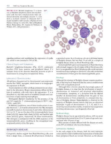
FIGURE 22.13 Burkitt’s ly pho . T is cl ssic
c se o Burkitt’s ly pho shows inter e i te-
size cells th t h ve roun e nuclei with con-
ense chro tin n ultiple nucleoli. T e cells
show o er te ount o cytopl s th t is
eeply b sophilic with v cuoles. (Reprinte ro
Pereir I, George I, Arber DA. Atlas o Peripheral
Blood, Phil elphi , PA: Lippincott Willi s &
Wilkins, 2011, with per ission.)
sign ling p thw y n o ul ting the expression o cyclin ger in l center. Reed-Sternberg cells re ef ning e ture
D3, which is lso ut te in 30% o BL. o Ho gkin ise se, but less th n 1% o cells in s ple o
Ho gkin ise se tissue re Ree -Sternberg cells.
Clinical Signs and Sym ptom s I unoglobulin gene re rr nge ents in Ree -Sternberg
Burkitt’s ly pho /leuke i (Fig. 22.13) co only cells strongly suggest B-cell origin o Ree -Sternberg cells,
involves CNS, bone rrow, n peripher l bloo . T e but it is i gin ble th t Ree -Sternberg cells coul represent
en e ic or o the isor er cl ssic lly presents s j w or tr ns or e croph ges or en ritic cells with no lous
ci l sses in young boys in equ tori l A ric . reco bin tion o their ger line i unoglobulin genes.
Etiology
Laboratory Characteristics
Although the etiology o Ho gkin ise se re ins question-
All subtypes ch r cterize by chro oso l re rr nge ents
involve the CMYC oncogene, which le s to its in ppropri- ble, it h s long been suspecte th t the c use is n in ectious
te expression in B cells. gent with long l tent perio .
ouch i prints or other cytologic prep r tions re ex - Although little is known bout the k ryotypic p ttern o
ine in the l bor tory. Biopsy e onstr tes i use inf ltr - Ho gkin ise se, it is cle r th t the involve ent o specif c
tion o neopl stic cells with st rry sky ppe r nce. T is is chro oso es in nu eric l n structur l bnor lities
escribe s sky blue nuclei o neopl stic ly phs n sc t- is nonr n o . Aneuploidy, or evi tion ro the iploi
tere st rs o p le-st ining croph ges. nu ber o chro oso es, resulting ro the g in or loss
Distinguish Burkitt’s ly pho by the presence o sIg o chro oso es or ro polyploi s, is ch r cteristic e -
n neg tivity. T e i unophenotypes is CD19+, sIg+, ture o Ho gkin ise se. Hyper iploi y is observe in the
CD10+, n CD5-. jority o Ho gkin ise se tu ors th t h ve n bnor l
In Burkitt’s ly pho , one o three tr nsloc tions is usu- k ryotype. A g in o chro oso es 1, 2, 5, 12, n 21 is
lly observe . T ese tr nsloc tions involve the Myc oncogene recurring nu eric l bnor lity; structur l re rr nge ents
nor lly loc te on chro oso e 8 n either chro oso e involving chro oso e 1 re requently observe .
14, 2, or 22. T ese re the sites o the i unoglobulin he vy
ch in [t(8;14)], k pp light ch in [t(2;8)], n l b light Epidem iology
ch in [t(8;22)] genes, respectively. In these tr nsloc tions, Ho gkin ise se h s n ge-rel te inci ence, with one pe k
Myc oncogene is juxt pose with the DNA sequence o the occurring in the perio ro 25 to 35 ye rs o ge n sec-
i unoglobulin genes, resulting in the unregul te tr n- on pe k er 50 ye rs o ge.
scription l ctivity o the Myc gene. Sixty percent o ults icte with the ise se re le,
s re 80% o chil ren who h ve the ise se.
HODGKIN DISEASE Laboratory Findings
In the e rly st ges o the ise se, both the tot l leukocyte
Cytogenetic stu ies suggest th t Ree -Sternberg cells rise count n the result o i erenti l ex in tion o leukocytes
ro single clone, co on B-cell precursor loc te in ro peripher l bloo re nor l. However, s the ise se