Page 510 - Clinical Hematology_ Theory _ Procedures ( PDFDrive )
P. 510

494            PART 7  ■  Principles and Disorders of Hemostasis and Thrombosis


                                                                                                                                                         Developmental Characteristics of
                                                                                                                                TABLE        25.4
                                                                                                                                                         Mature Megakaryocytic Cells


                                                                                                                                                              Megakaryocyte                                  Platelet


                                                                                                                               Size                           30–160 µm                                      2–4 µm


                                                                                                                               Nuclear-                       1:1–1:12

                                                                                                                               cytoplasmic ratio

                                                                                                                               Nucleus


                                                                                                                               Shape                          Lobulated (two or more                         (Anuclear)

                                                                                                                                                              lobes)

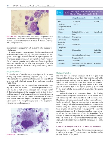
               FIGURE  25.4  Meg  k  ryobl  st  (see  arrow).  (Reprinte     ro                                                Chromatin color                Blue-purple                                    —

               An  erson SC. Anderson’s Atlas o  Hematology, Phil    elphi  , PA:

               Wolters Kluwer He  lth/Lippincott Willi    s & Wilkins, Copyright                                               Chromatin                      Granular                                       —

               2003, with per  ission.)                                                                                        clumping
                                                                                                                               Nucleoli                       Not visible                                    —


                ost pri itive progenitor cell co   itte   to   eg  k  ryo-                                                     Cytoplasm
               cyte line  ge.                                                                                                  Color                          Pinkish blue                                   Light-blue


                    T e next st  ge o    eg  k  ryocyte   evelop  ent is    s    ll,                                                                                                                         fragments
                 ononucle  r     rrow cell (Fig. 25.4) th  t expresses pl  telet-                                              Shape                          Occasional pseudopods


               specif c phenotypic     rkers but is not   orphologic  lly i  en-
               tif   ble   s      eg  k  ryocyte. T ese tr  nsition  l cells represent                                                                        Irregular border


               5% o      rrow   eg  k  ryocyte ele  ents. So  e tr  nsition  l                                                 Amount                         Abundant
               i      ture    eg  k  ryocyte  cells      y  be  c  p  ble  o   cellul  r                                       Granules                       Abundant near the borders  Scattered


                 ivision, but   ost   re nonproli er  ting while   ctively un  er-                                                                            of the cytoplasm
               going en  o  itosis.


               Megakaryocytes
                                                                                                                           Mature Platelets
               T e f n  l st  ge o    eg  k  ryocyte   evelop  ent is the   or-
               phologic  lly  i  entif   ble    eg  k  ryocyte  (Fig.  25.5).  T ese                                       Pl  telets  h  ve    n    ver  ge    i    eter  o   2  to  4  µ  ,  with


               cells   re re    ily recogniz  ble in the     rrow bec  use o  their                                        younger pl  telets being l  rger th  n ol  er ones. In contr  st to
               l  rge  size    n    lobul  te    nuclei.  T ese  cells    re  polyploi                                       eg  k  ryocytes, pl  telets h  ve no nucleus. T e cytopl  s   is


               (    ble 25.4).                                                                                             light blue, with evenly   isperse  , f ne re  -purple gr  nules.
                    Meg  k  ryocytes   re the l  rgest bone     rrow cells, r  ng-                                         An  in  ctive  or  unsti  ul  te    pl  telet  circul  tes    s      thin,


               ing  up  to  160  µ    in  size.  T e  nucle  r-cytopl  s  ic  (N:C)                                        s  ooth-sur   ce      isc.  T is    iscoi    sh  pe  is      int  ine
               r  tio c  n be   s high   s 1:12. Nucleoli   re no longer visible.                                          by the   icrotubul  r cytoskeleton bene  th the cytopl  s  ic


               A   istinctive  e  ture o  the   eg  k  ryocyte is th  t it is not                                            e  br  ne.
                ultinucle  te  . T e  ully     ture lobes o  the   eg  k  ryocyte                                               Pl  telets  circul  te  in    n  in  ctiv  te     or    in  the    owing


               she   pl  telets  ro   the cytopl  s   on co  pletion o      tur  -                                         bloo  stre     through en  otheliu  -line   bloo   vessels with-
               tion. Pl  telet  or    tion begins with the initi  l   ppe  r  nce o                                        out  inter  cting  with  other  pl  telets  or  with  the  vessel  w  ll.


                  pink color in the b  sophilic cytopl  s   o  the   eg  k  ryo-                                           Pl  telets   re extre  ely sensitive cells   n       y respon   to   in-
               cyte   n   incre  se   gr  nul  rity.                                                                       i    l sti  ul  tion by  or  ing pseu  opo  s th  t spont  neously
                                                                                                                           retr  ct. Stronger sti  ul  tion c  uses pl  telets to beco  e sticky

                                                                                                                           without losing their   iscoi   sh  pe; however, ch  nges in sh  pe

                                                                                                                           to   n irregul  r sphere with spiny pseu  opo  s will occur with

                                                                                                                                ition l sti  ul  tion. T is  lter  tion in cellul  r sh  pe is trig-

                                                                                                                           gere   by   n incre  se in the level o  cytopl  s  ic c  lciu  . Such

                                                                                                                           ch  nges in sh  pe   cco  p  nie   by intern  l cellul  r contr  c-

                                                                                                                           tions c  n result in the rele  se o      ny o  the intern  l org  n-

                                                                                                                           elles. A loss o  vi  bility is   ssoci  te   with this ch  nge to    spiny

                                                                                                                           sphere.


                                                                                                                           Cellular Ultrastructure of a Mature Platelet


                                                                                                                           Ex    in  tion o     pl  telet with   n electron   icroscope reve  ls

                                                                                                                             v  riety o  structures. T ese structures   re  un      ent  l to

               FIGURE 25.5  Meg  k  ryocyte.                                                                               the  unctioning o  the pl  telet.
   505   506   507   508   509   510   511   512   513   514   515