Page 706 - Clinical Hematology_ Theory _ Procedures ( PDFDrive )
P. 706

690            PART 8  ■  Fundamentals of Hematological Analysis


                       SPECIAL STAINS (continued)


               Di  use staining may be present in more mature nucleate                                                     SIDEROCYTE STAIN: PRUSSIAN BLUE STAINING

               erythrocytes.                                                                                               METHOD

                    T is proce  ure in CLSI  ormat is provi  e   on this book’s                                            Principle

               companion Web site at thepoint.lww.com/  urgeon6e.
                                                                                                                           Te Prussian blue reaction precipitates  ree iron into small blue


               PEROXIDASE (MYELOPEROXIDASE) IN                                                                             or blue-green granules in erythrocytes. Free iron is not i  enti-

               LEUKOCYTES: CYTOCHEMICAL STAINING                                                                           fable on Wright- or Wright-Giemsa–staine   bloo   smears. An

               METHOD                                                                                                      immature or mature erythrocyte containing  ree iron is re erre

               Principle                                                                                                   to as a si  eroblast or si  erocyte, respectively. Increase   numbers
                                                                                                                           o  si  erocytes are seen in   isor  ers such as thalassemia major or
               Myeloperoxi  ase (MP) is   etecte   by means o  the enzyme’s                                                in patients a  er a splenectomy. I  the iron granules encircle the

               interaction with   iaminobenzi  ine (DAB), a benzi  ine substi-                                             nucleus o  the erythrocyte, it is re erre   to as a ringe   si  eroblast.

               tute. T e brown reaction pro  uct is f rst intensif e   with copper                                         Although alcoholism is the most common cause o  ringe   si  ero-

               salts  ollowe   by Gill’s mo  if e   Papanicolaou stain, which results                                      blasts, they may also be seen in cases o  lea   poisoning or anemia.

               in intense gray-black granules at sites o  neutrophil an   mono-

               cyte MP activity (Fig. 32.17). T e reaction can be illustrate   as:                                         Clinical Applications

                                                                                                                           An increase in si  eroblasts is associate   with thalassemia major
                       DAB H O                    MP     →    oxi  ize  DAB
                                +
                                       2   2                                                                               or minor an   with the si  eroblastic anemias. T e si  eroblastic
                                                              ( light brown pigment)                                       anemias are a miscellaneous group o    iseases cause   by   rugs

                                                         (  )
                       Oxi  ize  DAB Cu NO  → gray-black pigment                                                           or chemicals, as well as various   isor  ers, an   are o  here  itary
                                                +
                                                                 33
                                                                    2
                                                                                                                           or i  iopathic origin. Si  erocytes are uncommon in peripheral
                    Tis proce  ure   i  erentiates cells o  lymphoi   origin  rom                                          bloo   but may be seen a  er a splenectomy.

               granulocytes an   their precursors an   monocytes.                                                               Tis proce  ure in CLSI  ormat is provi  e   on this book’s


               Clinical Applications                                                                                       companion Web site at thepoint.lww.com/  urgeon6e.


               T e most probable results to be expecte   with peroxi  ase-                                                 SUDAN BLACK B STAIN: CYTOCHEMICAL

               staining proce  ures in cases o  acute nonlymphocytic leuke-                                                STAINING METHOD

               mia (ANLL) are:                                                                                             Principle


               M1 →5% to 15% o  blasts may be positive                                                                     Following f xation, bloo   or bone marrow f lms are immerse
               M2 an   M3 →positive                                                                                        in a bu  ere   Su  an black B solution. A  er rinsing, sli  es are


               M4 →positive (usually a mixe   population o  cells)                                                         counterstaine   with Mayer’s hematoxylin. Cells are examine

               M5a →may be positive                                                                                        microscopically  or the presence o  blue-black   iscrete gran-
               M5b →a f ne granular   eposit may be observe   in more                                                      ulation (Fig. 32.18). Cells committe   to the lymphoi   path-

                      mature cells                                                                                         way   isplay negative staining reactions, whereas myeloi   an

               M6 an   M7 →myelocytic cells are positive                                                                   monocytoi    orms   isplay characteristic positive  reactions.

               ALL →negative (L1, L2, an   L3)                                                                             Te Su  an black B staining pattern usually parallels the MP

                                                                                                                           stain an   is use ul in the i  entif cation o  myelogenous an
                    Tis proce  ure in CLSI  ormat is provi  e   on this book’s

               companion Web site at thepoint.lww.com/  urgeon6e.                                                          myelomonocytic leukemias.


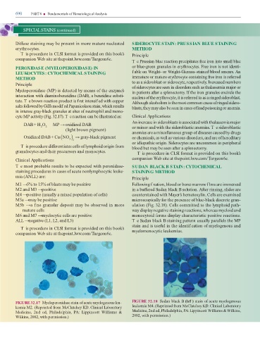
               FIGURE 32.17  Myeloperoxi  ase stain o  acute myelogenous leu-                                              FIGURE 32.18  Su  an black B (lef ) stain o  acute myelogenous

               kemia M2. (Reprinte    rom McClatchey KD. Clinical Laboratory                                               leukemia M4. (Reprinte    rom McClatchey KD. Clinical Laboratory
               Medicine,  2n    e  ,  Phila  elphia,  PA:  Lippincott  Williams  &                                         Medicine, 2n   e  , Phila  elphia, PA: Lippincott Williams & Wilkins,

               Wilkins, 2002, with permission.)                                                                            2002, with permission.)
   701   702   703   704   705   706   707   708   709   710   711