
Page 705 - Clinical Hematology_ Theory _ Procedures ( PDFDrive )
P. 705
CHAPTER 32 ■ Laboratory Manual: Manual Procedures in Hematology 689
SPECIAL STAINS (continued)
Clinical Applications bloo . T ese cells shoul appear rarely. T e results are
Heinz bo ies are orme when the glycolytic enzymes in the expresse as a percentage. Normal a ults have less than 1%
erythrocytes are unable to prevent the oxi ation o hemo- Hb F–containing cells. In ant values are higher, with new-
globin. As a result, the hemoglobin is eventually enature born in ants having 70% to 90% Hb F–containing cells.
an precipitate to orm Heinz bo ies. Erythrocytic enzyme Clinical Applications
systems ecrease as the cell ages; there ore, occasional Heinz
bo ies will be observe in normal bloo . Increase amounts o Hb F are oun in various hemoglobin-
Increase numbers o Heinz bo ies represent unstable opathies such as here itary persistence o etal hemoglobin,
orms o hemoglobin that are present in a number o hemo- sickle cell anemia, an the thalassemias.
lytic isor ers. Heinz bo ies occur in isor ers such as G6PD T is proce ure in CLSI ormat is provi e on this book’s
or glutathione ef ciencies, secon ary to the action o certain companion Web site at thepoint.lww.com/ urgeon6e.
oxi ant rugs, an in the presence o unstable hemoglobins PERIODIC ACID-SCHIFF (PAS) IN LEUKOCYTES:
such as Hb Zurich an Hb H. CYTOCHEMICAL OR HISTOCHEMICAL STAINING
T is proce ure in CLSI ormat is provi e on this book’s METHOD
companion Web site at thepoint.lww.com/ urgeon6e.
Principle
HEMOGLOBIN F DETERMINATION BY ACID When treate with perio ic aci , glycols are oxi ize to
ELUTION: KLEIHAUER AND BETKE METHOD al ehy es. A er reaction with Schi ’s reagent (a mixture o
MODIFIED BY SHEPARD, WEATHERALL, AND pararosaniline an so ium metabisulf te), a pararosaniline
CONLEY a uct is release that stains the glyco-containing cellular
Principle elements (Fig. 32.16). Clinically, the PAS stain is help ul in
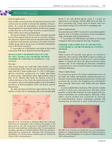
A er bloo smears are f xe with ethyl alcohol, a citric recognizing some cases o erythroleukemia an acute lym-
aci ›phosphate bu er solution removes (elutes) hemoglobin phoblastic leukemia.
other than Hb F rom erythrocytes. T e Hb F ( etal hemo- Clinical Applications
globin)–containing erythrocytes are visibly i enti iable In normal bone marrow, the earliest myeloi precursors o
on microscopic examination when appropriately staine not stain, but i use an granular staining increases as a
(Fig. 32.15). Shortly a er birth, the amount o Hb F in unction o maturation along myeloi pathways. Erythrocytic
humans ecreases to low levels. Increase amounts o Hb F precursors o not stain, but megakaryocytes an platelets
are oun in various hemoglobinopathies such as here itary stain intensely. Monocytes stain aintly an may isplay
persistence o etal hemoglobin, sickle cell anemia, an the granules.
thalassemias. In acute lymphoblastic leukemia, PAS activity is highly
An a ult specimen shoul have approximately the same variable. In most cases, some precursor cells show coarse
number o ense Hb F–containing cells as the normal a ult
granules or block-like positivity. In acute granulocytic leu-
kemia, myeloblasts are usually negative, although a aint
i use reaction pro uct may occasionally be observe .
In erythroleukemia, intense cytoplasmic granular PAS
staining may be observe in early erythroi precursors.
Adult
Fetal
cell cell
FIGURE 32.15 Aci elution or Kleihauer-Betke stain. Cell type:
Re bloo cell. Description: Cells containing hemoglobin F will
appear pink to re ; cells containing no hemoglobin F will only
have their outer membrane visible (ghost cells). Clinical conditions:
Here itary persistence o etal hemoglobin MDS some leukemias. FIGURE 32.16 Perio ic aci -Schi stain o acute lymphoblastic
(Reprinte rom An erson SC. Anderson’s Atlas of Hematology, leukemia L1. (Reprinte rom McClatchey KD. Clinical Laboratory
Phila elphia, PA: Wolters Kluwer Health/Lippincott Williams & Medicine, 2n e , Phila elphia, PA: Lippincott Williams & Wilkins,
Wilkins, 2003, with permission.) 2002, with permission.)
(continued)