
Page 221 - Review of Medical Microbiology and Immunology ( PDFDrive )
P. 221
mebooksfree.com
mebooksfree.com
mebooksfree.com
mebooksfree.com
mebooksfree.com
mebooksfree.com
mebooksfree.com mebooksfree.com mebooksfree.com pneumonia 2 to 12 weeks after birth. Chlamydial conjunc- mebooksfree.com
mebooksfree.com
mebooksfree.com
PART II Clinical Bacteriology
210
infected primarily by inhaling organisms in airborne dry
bird feces.
tivitis also occurs in adults as a result of the transfer of
Sexually transmitted disease caused by C. trachomatis
organisms from the genitals to the eye.
Patients with genital tract infections caused by C. tra-
occurs worldwide, but trachoma is most frequently found
chomatis have a high incidence of reactive arthritis and
in developing countries in dry, hot regions such as north-
ern Africa. Trachoma is a leading cause of blindness in
those countries.
arthritis, and uveitis. These are autoimmune diseases
caused by antibodies formed against C. trachomatis cross-
Patients with a sexually transmitted disease are coin-
reacting with antigens on the cells of the urethra, joints,
fected with both C. trachomatis and Neisseria gonorrhoeae Reiter’s syndrome, which is characterized by urethritis,
mebooksfree.com
and uveal tract (see Chapter 66).
in approximately 10% to 30% of cases.
mebooksfree.com mebooksfree.com mebooksfree.com with lesions on genitalia and in lymph nodes. mebooksfree.com mebooksfree.com
Chlamydia trachomatis L1–L3 immunotypes cause lym-
phogranuloma venereum, a sexually transmitted disease
Pathogenesis & Clinical Findings
Chlamydiae infect primarily epithelial cells of the mucous
Infection by C. trachomatis leads to formation of anti-
membranes or the lungs. They rarely cause invasive, dis-
bodies and cell-mediated reactions but not to resistance to
seminated infections.
reinfection or elimination of organisms.
CHLAMYDIA TRACHOMATIS
CHLAMYDIA PNEUMONIAE
Chlamydia trachomatis has more than 15 immunotypes
(A–L). Types A, B, and C cause trachoma, a chronic con-
tract infections, especially bronchitis and pneumonia, in
junctivitis endemic in Africa and Asia. Trachoma may
young adults. Most infections are mild or asymptomatic.
recur over many years and may lead to blindness but causes Chlamydia pneumoniae causes upper and lower respiratory
The clinical picture resembles other atypical pneumonias,
mebooksfree.com
mebooksfree.com mebooksfree.com mebooksfree.com CHLAMYDIA PSITTACI mebooksfree.com mebooksfree.com
no systemic illness.
especially that caused by Mycoplasma pneumoniae. It is
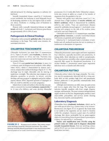
Types D–K cause genital tract infections. In men, it is
unclear whether C. pneumoniae causes upper respiratory
a common cause of nongonococcal urethritis (often abbre-
infections such as sinusitis and otitis media.
viated NGU), which is characterized by dysuria and a
watery, nonpurulent urethral discharge (Figure 25–3). The
discharge may be slight, detectable only by staining of
Chlamydia psittaci infects the lungs primarily. The infec-
underwear overnight. This infection may progress to epi-
didymitis, prostatitis, or proctitis. In women, cervicitis
tion may be asymptomatic (detected only by a rising anti-
body titer) or may produce high fever and pneumonia.
develops and may progress to salpingitis and pelvic inflam-
matory disease (PID). Repeated episodes of salpingitis or
human to human.
PID can result in infertility or ectopic pregnancy.
Although most infections are mild, some are quite severe
Infants born to infected mothers often develop mucopu-
rulent conjunctivitis (neonatal inclusion conjunctivitis) 7 Human psittacosis is not generally communicable from
and involve organs other than the lung. The respiratory
mebooksfree.com mebooksfree.com mebooksfree.com (myocarditis, pericarditis), and nervous system (hearing mebooksfree.com
mebooksfree.com
mebooksfree.com
to 12 days after delivery, and some develop chlamydial
infection typically manifests with fever, cough, dyspnea,
myalgias, and headache. The most common extrapulmonary
organs involved are the liver (hepatomegaly, jaundice), heart
loss, transverse myelitis, and encephalitis).
Laboratory Diagnosis
Chlamydiae form cytoplasmic inclusions, which can be
seen with special stains (e.g., Giemsa stain) or by immuno-
fluorescence (see Figure 25–2). In general, the Gram stain
is not useful as the organisms are too small to visualize
within the cytoplasm. However, a gram stain of a urethral
discharge that shows neutrophils but no gram-negative
diplococci resembling Neisseria gonorrhoeae is presumptive
mebooksfree.com mebooksfree.com mebooksfree.com patient’s urine are widely used to diagnose chlamydial mebooksfree.com
mebooksfree.com
mebooksfree.com
evidence for infection by C. trachomatis.
Nucleic acid amplification tests (NAATs) using the
FIGURE 25–3
Nongonococcal urethritis. Note watery, nonpu-
sexually transmitted disease. Tests not involving culture,
rulent discharge caused by Chlamydia trachomatis. The urethral
such as NAAT, are now more commonly used than culture-
discharge caused by Neisseria gonorrhoeae is more mucoid and purulent.
based tests (see later).
(Used with permission from Seattle STD/HIV Prevention Training Center.)
mebooksfree.com mebooksfree.com mebooksfree.com mebooksfree.com mebooksfree.com mebooksfree.com