
Page 220 - Review of Medical Microbiology and Immunology ( PDFDrive )
P. 220
mebooksfree.com
mebooksfree.com
mebooksfree.com
mebooksfree.com
mebooksfree.com
mebooksfree.com
mebooksfree.com mebooksfree.com Extracellular infectious Attachment and mebooksfree.com mebooksfree.com mebooksfree.com
mebooksfree.com
CHAPTER 25 Chlamydiae
209
entry of elementary body
Cell nucleus
elementary body
Formation of
reticulate body
Release
mebooksfree.com mebooksfree.com Elementary bodies mebooksfree.com mebooksfree.com mebooksfree.com
mebooksfree.com
Multiplication of reticulate
Multiplication
bodies by binary fission
ceases
Reticulate bodies
Development of a
Reorganization of
large cytoplasmic
reticulate bodies into elementary bodies
inclusion
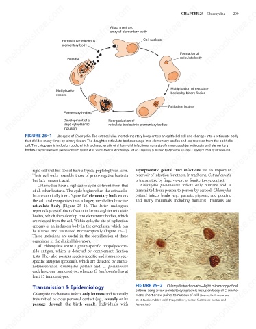
FIGURE 25–1 Life cycle of Chlamydia. The extracellular, inert elementary body enters an epithelial cell and changes into a reticulate body
mebooksfree.com
mebooksfree.com
mebooksfree.com mebooksfree.com mebooksfree.com asymptomatic genital tract infections are an important mebooksfree.com
that divides many times by binary fission. The daughter reticulate bodies change into elementary bodies and are released from the epithelial
cell. The cytoplasmic inclusion body, which is characteristic of chlamydial infections, consists of many daughter reticulate and elementary
bodies. (Reproduced with permission from Ryan K et al. Sherris Medical Microbiology. 3rd ed. Originally published by Appleton & Lange. Copyright 1994 by McGraw-Hill.)
rigid cell wall but do not have a typical peptidoglycan layer.
reservoir of infection for others. In trachoma, C. trachomatis
Their cell walls resemble those of gram-negative bacteria
is transmitted by finger-to-eye or fomite-to-eye contact.
but lack muramic acid.
Chlamydia pneumoniae infects only humans and is
Chlamydiae have a replicative cycle different from that
of all other bacteria. The cycle begins when the extracellu-
psittaci infects birds (e.g., parrots, pigeons, and poultry,
lar, metabolically inert, “sporelike” elementary body enters
and many mammals including humans). Humans are
the cell and reorganizes into a larger, metabolically active transmitted from person to person by aerosol. Chlamydia
reticulate body (Figure 25–1). The latter undergoes
mebooksfree.com mebooksfree.com mebooksfree.com mebooksfree.com mebooksfree.com mebooksfree.com
repeated cycles of binary fission to form daughter reticulate
bodies, which then develop into elementary bodies, which
are released from the cell. Within cells, the site of replication
appears as an inclusion body in the cytoplasm, which can
be stained and visualized microscopically (Figure 25–2).
These inclusions are useful in the identification of these
organisms in the clinical laboratory.
All chlamydiae share a group-specific lipopolysaccha-
ride antigen, which is detected by complement fixation
tests. They also possess species-specific and immunotype-
specific antigens (proteins), which are detected by immu-
nofluorescence. Chlamydia psittaci and C. pneumoniae
each have one immunotype, whereas C. trachomatis has at
mebooksfree.com
mebooksfree.com mebooksfree.com mebooksfree.com FIGURE 25–2 Chlamydia trachomatis—light microscopy of cell mebooksfree.com
mebooksfree.com
least 15 immunotypes.
Transmission & Epidemiology
culture. Long arrow points to cytoplasmic inclusion body of C. tracho-
Chlamydia trachomatis infects only humans and is usually
matis; short arrow points to nucleus of cell. (Source: Dr. E. Arum and
transmitted by close personal contact (e.g., sexually or by
Dr. N. Jacobs, Public Health Image Library, Centers for Disease Control and
passage through the birth canal). Individuals with
Prevention.)
mebooksfree.com mebooksfree.com mebooksfree.com mebooksfree.com mebooksfree.com mebooksfree.com