
Page 528 - Review of Medical Microbiology and Immunology ( PDFDrive )
P. 528
mebooksfree.com
mebooksfree.com
mebooksfree.com
mebooksfree.com
mebooksfree.com mebooksfree.com mebooksfree.com adjacent IgE molecules are cross-linked by antigen, immu- 517 mebooksfree.com
mebooksfree.com
mebooksfree.com
mebooksfree.com
mebooksfree.com
CHAPTER 58 Cellular Basis of the Immune Response
larvae is mediated by receptors on the eosinophil surface
for the Fc portion of the heavy chain of IgG and IgE.
nologically active mediators, such as histamine, and
Another function of eosinophils may be to mitigate the
enzymes, such as peroxidases and hydrolases, are released.
These cause inflammation and, when produced in large
effects of immediate hypersensitivity reactions because the
granules of eosinophils contain histaminase, an enzyme
amounts, cause severe immediate hypersensitivity reac-
that degrades histamine, which is an important mediator of
Mast cells also play an important role in the innate
immediate reactions. However, the granules of the eosino-
phils also contain leukotrienes and peroxidases, which can
response to bacteria and viruses. The surface of mast cells
contains Toll-like receptors that recognize bacteria and
damage tissue and cause inflammation. The granules also tions such as systemic anaphylaxis.
mebooksfree.com mebooksfree.com mebooksfree.com infection. Dendritic cells are important APCs that initiate mebooksfree.com
viruses. The mast cells respond by releasing cytokines and
contain major basic protein that damages respiratory epi-
mebooksfree.com
mebooksfree.com
enzymes from their granules that mediate inflammation
thelium and contributes to the pathogenesis of asthma.
and attract neutrophils and dendritic cells to the site of
Eosinophils can phagocytose bacteria but they do so
weakly and are not sufficient to protect against pyogenic bac-
the adaptive response. The role of mast cells in inflamma-
terial infections in neutropenic patients. Although they can
phagocytose, they do not present antigen to helper T cells.
tion has been demonstrated in rheumatoid arthritis. These
The growth and differentiation of eosinophils are stimulated
cells produce both inflammatory cytokines and the
by IL-5. Eotaxin is a chemokine that attracts eosinophils to
enzymes that degrade the cartilage in the joints.
the site of nematode larvae and allergic reactions.
BASOPHILS & MAST CELLS
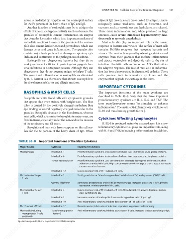
The important functions of the main cytokines are
described in Table 58–8. Note that the three important
Basophils are white blood cells with cytoplasmic granules IMPORTANT CYTOKINES
proinflammatory cytokines are IL-1, IL-6, and TNF. The
mebooksfree.com
mebooksfree.com mebooksfree.com mebooksfree.com IL-10 and transforming growth factor β. mebooksfree.com mebooksfree.com
that appear blue when stained with Wright stain. The blue
term proinflammatory means “to stimulate or enhance
color is caused by the positively charged methylene blue
inflammation.” The main anti-inflammatory cytokines are
dye binding to several negatively charged molecules in the
granules. Basophils circulate in the bloodstream, whereas
mast cells, which are similar to basophils in many ways, are
Cytokines Affecting Lymphocytes
fixed in tissue, especially under the skin and in the mucosa
(1) IL-1 is produced mainly by macrophages. It is a pro-
of the respiratory and GI tracts.
inflammatory cytokine (i.e., plays an important role, along
Basophils and mast cells have receptors on the cell sur-
with IL-6 and TNF, in inducing inflammation). In addition,
face for the Fc portion of the heavy chain of IgE. When
TABLE 58–8 Important Functions of the Main Cytokines
Major Source Cytokine Important Functions
mebooksfree.com
mebooksfree.com mebooksfree.com Tumor necrosis factor Proinflammatory cytokine. Low concentration: activates neutrophils and increases their mebooksfree.com
mebooksfree.com
mebooksfree.com
Interleukin-1
Proinflammatory cytokine. Induces fever. Induces liver to produce acute-phase proteins.
Macrophages
Proinflammatory cytokine. Induces fever. Induces liver to produce acute-phase proteins.
Interleukin-6
adhesion to endothelial cells. High concentration: mediates septic shock, acts as cachectin,
causes necrosis of tumors.
Drives development of Th-1 subset of T cells.
Interleukin-12
T-cell growth factor. Stimulates growth of both helper (CD4) and cytotoxic (CD8) T cells.
Th-1 subset of helper
Interleukin-2
T cells
Stimulates phagocytosis and killing by macrophages. Increases class I and II MHC protein
Gamma interferon
expression. Inhibits growth of Th-2 cells.
Drives development of Th-2 subset of T cells. Stimulates B-cell growth. Increases isotype
Interleukin-4
Th-2 subset of helper
class switching to IgE.
T cells
mebooksfree.com mebooksfree.com Interleukin-5 Increases number of eosinophils. Increases isotype class switching to IgA. mebooksfree.com mebooksfree.com
mebooksfree.com
mebooksfree.com
Interleukin-10
Anti-inflammatory cytokine. Inhibits development of Th1 subset of T cells.
Recruits neutrophils to site of infection. Important in gut mucosal immunity.
Th-17 subset of T cells
Interleukin-17
Many cells including
Transforming growth
Anti-inflammatory cytokine. Inhibits activation of T cells. Increases isotype switching to IgA.
factor-β
macrophages, T cells,
and B cells
Ig = immunoglobulin; MHC = major histocompatibility complex.
mebooksfree.com mebooksfree.com mebooksfree.com mebooksfree.com mebooksfree.com mebooksfree.com