


Page 477 - Textbook of Pathology, 6th Edition
P. 477
461


            Chapter 17
            Chapter 17                         The Respiratory System


                                LUNGS


           NORMAL STRUCTURE
           ANATOMY. The normal adult right lung weighs 375 to 550
           gm (average 450 gm) and is divided by two fissures into three
           lobes—the upper, middle and lower lobes. The weight of the
           normal adult left lung is 325 to 450 gm (average 400 gm) and
           has one fissure dividing it into two lobes—the upper and
           lower lobes, while the middle lobe is represented by the
           lingula. The airways of the lungs arise from the trachea by
           its division into right and left main bronchi which continue
           to divide and subdivide further, eventually terminating into
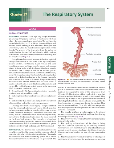
           the alveolar sacs (Fig. 17.1).
              The right main bronchus is more vertical so that aspirated
           foreign material tends to pass down to the right lung rather
           than to the left. The trachea, major bronchi and their
           branchings possess cartilage, smooth muscle and mucous                                                     CHAPTER 17
           glands in their walls, while the bronchioles have smooth
           muscle but lack cartilage as well as the mucous glands.
           Between the tracheal bifurcation and the smallest bronchi,
           about 8 divisions take place. The bronchioles so formed further
           undergo 3 to 4 divisions leading to the terminal bronchioles
           which are less than 2 mm in diameter. The part of the lung  Figure 17.1  The structure of an acinus which is part of the lung
           tissue distal to a terminal bronchiole is called an acinus. A  distal to terminal bronchiole. It shows respiratory bronchiole, alveolar
           cluster of about 5 acini supplied by terminal bronchioles and  ducts and alveolar sacs.
           enclosed by visible fibrous septa is termed as the pulmonary  mucosa of bronchi contains numerous submucosal mucous
           lobule. An acinus consists of 3 parts:                                                                     The Respiratory System
                                                               glands and neuroendocrine cells which are bronchial counter-
           1. Several (usually 3 to 5 generations) respiratory bronchioles  parts of the argentaffin cells of the alimentary tract
           originate from a terminal bronchiole.
                                                               (Chapter 20). The structure of bronchioles differs from that
           2. Each respiratory bronchiole divides into several alveolar  of bronchi and its subdivisions as well as from alveoli. They
           ducts.                                              are lined by a single layer of pseudostratified columnar
           3. Each alveolar duct opens into many alveolar sacs (alveoli)  ciliated epithelium but no mucus cells and hence, unlike the
           which are blind ends of the respiratory passages.   bronchi, contain no mucus secretion on the surface. They
              The lungs have double blood supply—oxygenated blood  contain some nonciliated Clara cells which secrete protein
           from the bronchial arteries and venous blood from the  rich in lysozyme and immunoglobulins but unlike the alveoli
           pulmonary arteries, and there is mixing of the blood to some  contain no surfactant.
           extent. In case of blockage of one side of circulation, the  The alveolar walls or alveolar septa are the sites of
           supply from the other can maintain the vitality of pulmonary  exchange between the blood and air and have the following
           parenchyma. The bronchial veins drain the blood supplied  microscopic features (Fig. 17.2):
           by the bronchial arteries. The lungs have abundant  1. The capillary endothelium lines the anastomotic capillaries
           intercommunicating lymphatics on the surface which drain  in the alveolar walls.
           into the subpleural plexus. Hilar and tracheobronchial lymph  2. The capillary endothelium and the, alveolar lining
           nodes receive the lymph and drain into the thoracic duct.  epithelial cells are separated by the  capillary basement
                                                               membrane and some interstitial tissue. The interstitial tissue
           HISTOLOGY. The bronchi and their subdivisions up to  consists of scanty amount of collagen, fibroblasts, fine elastic
           bronchioles are lined by pseudostratified columnar ciliated  fibres, smooth muscle cells, a few mast cells and mononuclear
           epithelial cells, also called respiratory epithelium. These cells  cells.
           are admixed with mucus-secreting goblet cells which  3. The alveolar epithelium consists of 2 types of cells: type I or
           decrease in number as the bronchioles are approached. The  membranous pneumocytes are the most numerous covering

