


Page 474 - Textbook of Pathology, 6th Edition
P. 474
458                                                      of acute pericarditis already described. Included under this
                                                               are: tuberculous pericarditis, chronic adhesive pericarditis,
                                                               chronic constrictive pericarditis, and the pericardial plaques.
                                                               1. TUBERCULOUS PERICARDITIS.  Tuberculous peri-
                                                               carditis is the most frequent form of granulomatous
                                                               inflammation of the pericardium. The lesions may occur by
                                                               one of the following mechanisms:
                                                               i) Direct extension from an adjacent focus of tuberculosis.
                                                               ii) By lymphatic spread e.g. from tracheobronchial lymph
                                                               nodes, chronic pulmonary tuberculosis or infected pleura.
                                                                  The exudate is slightly turbid, caseous or blood-stained
                                                               with sufficient fibrin. Tubercles are generally visible on the
                                                               pericardial surfaces and sometimes caseous areas are also
                                                               visible to the naked eye.

                                                                 Microscopically, typical tuberculous granulomas with
                                                                 caseation necrosis are seen in the pericardial wall. The
                                                                 lesions generally do not resolve but heal by fibrosis and
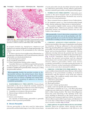
           Figure 16.34  Fibrinopurulent pericarditis. The pericardium is covered  calcification resulting in chronic constrictive pericarditis.
           with pink serofibrinous exudates. The space between the layers of the
           pericardium contains numerous inflammatory cells, chiefly PMNs.  2. CHRONIC ADHESIVE PERICARDITIS.  Chronic
                                                               adhesive pericarditis is the stage of organisation and healing
           by pyogenic bacteria (e.g. staphylococci, streptococci and  by formation of fibrous adhesions in the pericardium
           pneumococci) and less frequently by fungi and parasites. The  following preceding fibrinous, suppurative or haemorrhagic
           infection may spread to the pericardium by the following  pericarditis. The process begins by formation of granulation
           routes:                                             tissue and neovascularisation. Subsequently, fibrous
           i) By direct extension from neighbouring inflammation e.g.  adhesions develop between the parietal and the visceral
           in empyema of the pleural cavity, lobar pneumonia, infective  layers of the pericardium and obliterate the pericardial space
     SECTION III
           endocarditis and mediastinal infections.            (Fig. 16.35,A). Sometimes, fibrous adhesions develop
           ii) By haematogenous spread.                        between the parietal pericardium and the adjacent
           iii) By lymphatic permeation.                       mediastinum and is termed as adhesive mediastinopericarditis.
           iv) Direct implantation during cardiac surgery.     Chronic adhesive pericarditis differs from chronic
              Generally, fibrinous or serofibrinous pericarditis precedes  constrictive pericarditis in not embarrassing the function of
           the development of purulent pericarditis. The amount of  the heart. However, cardiac hypertrophy and dilatation may
           exudate is variable and is generally thick, creamy pus, coating  occur in severe cases due to increased workload.
           the pericardial surfaces.                           3. CHRONIC CONSTRICTIVE PERICARDITIS. This is
                                                               a rare condition characterised by dense fibrous or fibrocalcific
            Microscopically, besides the purulent exudate on the  thickening of the pericardium resulting in mechanical
            pericardial surfaces, the serosal layers show dense  interference with the function of the heart and reduced
     Systemic Pathology
            infiltration by neutrophils (Fig. 16.34). Purulent exudate  cardiac output. The condition usually results from a long-
            generally does not resolve completely but instead heals  standing preceding causes, e.g.
            by organisation resulting in adhesive or chronic   i) Tuberculous pericarditis
            constrictive pericarditis.                         ii) Purulent pericarditis
                                                               iii) Haemopericardium
           4. HAEMORRHAGIC PERICARDITIS. Haemorrhagic          iv) Concato’s disease (polyserositis)
           pericarditis is the one in which the exudate consists of  v) Rarely, acute non-specific and viral pericarditis.
           admixture of an inflammatory effusion of one of the foregoing  The heart is encased in 0.5 to 1 cm thick and dense
           types alongwith blood. The causes are as under:     collagenous scar which may be calcified. As a result, the heart
           i) Neoplastic involvement of the pericardium        fails to dilate during diastole. The dense fibrocollagenous
           ii) Haemorrhagic diathesis with effusion            tissue may cause narrowing of the openings of the vena
           iii) Tuberculosis                                   cavae, resulting in obstruction to the venous return to the
           iv) Severe acute infections                         right heart and consequent right heart failure. In contrast to
              The outcome of haemorrhagic pericarditis is generally  chronic adhesive pericarditis, hypertrophy and dilatation do
           similar to that of purulent pericarditis.           not occur due to dense fibrous scarring. Instead, the heart
                                                               size is normal or smaller (Fig. 16.35,B).
           B. Chronic Pericarditis
                                                               4. PERICARDIAL PLAQUES (MILK SPOTS, SOLDIERS’
           Chronic pericarditis is the term used for tuberculous  SPOTS).  These are opaque, white, shining and well-
           pericarditis and the healed stage of one of the various forms  circumscribed areas of organisation with fibrosis in the

