


Page 478 - Textbook of Pathology, 6th Edition
P. 478
462                                                      2. Pulmonary vascular disease
                                                               3. Pulmonary infections
                                                               4. Chronic obstructive pulomonary disease
                                                               5. Chronic restrictive pulmonary disease
                                                               6. Tumours of lungs.

                                                               PAEDIATRIC LUNG DISEASE
                                                               A number of congenital anomalies (e.g. agenesis, hypoplasia,
                                                               heterotopic tissue, vascular anomalies, tracheal and bronchial
                                                               anomalies, congenital pulmonary overinflation or lobar
                                                               emphysema, congenital cysts and bronchopulmonary
                                                               sequestration) and certain neonatal acquired lung diseases
                                                               (respiratory distress syndrome or hyaline membrane disease,
                                                               bronchopulmonary dysplasia, meconium aspiration
                                                               syndrome, persistent foetal circulation, atelactasis, collapse
                                                               and bronchiolitis) have been described. Some of the
                                                               important conditions from point of view of pathology are
                                                               discussed below.

                                                               CONGENITAL CYSTS

                                                               Developmental defects involving deficiency of bronchial or
                                                               bronchiolar cartilage, elastic tissue and muscle result in
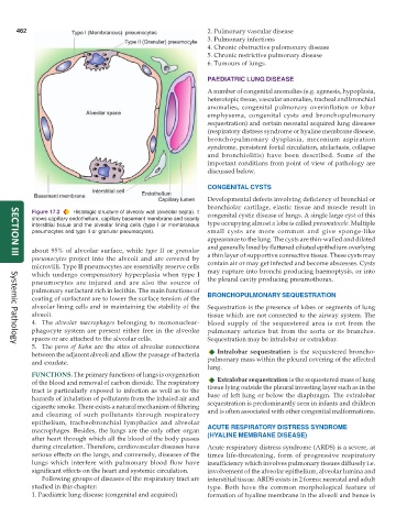
           Figure 17.2  Histologic structure of alveolar wall (alveolar septa). It  congenital cystic disease of lungs. A single large cyst of this
           shows capillary endothelium, capillary basement membrane and scanty
           interstitial tissue and the alveolar lining cells (type I or membranous  type occupying almost a lobe is called pneumatocele. Multiple
           pneumocytes and type II or granular pneumocytes).   small cysts are more common and give sponge-like
                                                               appearance to the lung. The cysts are thin-walled and dilated
                                                               and generally lined by flattened ciliated epithelium overlying
           about 95% of alveolar surface, while  type II or  granular  a thin layer of supportive connective tissue. These cysts may
           pneumocytes  project into the alveoli and are covered by
     SECTION III
           microvilli. Type II pneumocytes are essentially reserve cells  contain air or may get infected and become abscesses. Cysts
           which undergo compensatory hyperplasia when type I  may rupture into bronchi producing haemoptysis, or into
           pneumocytes are injured and are also the source of  the pleural cavity producing pneumothorax.
           pulmonary surfactant rich in lecithin. The main functions of
           coating of surfactant are to lower the surface tension of the  BRONCHOPULMONARY SEQUESTRATION
           alveolar lining cells and in maintaining the stability of the  Sequestration is the presence of lobes or segments of lung
           alveoli.                                            tissue which are not connected to the airway system. The
           4. The  alveolar macrophages belonging to mononuclear-  blood supply of the sequestered area is not from the
           phagocyte system are present either free in the alveolar  pulmonary arteries but from the aorta or its branches.
           spaces or are attached to the alveolar cells.       Sequestration may be intralobar or extralobar.
           5. The pores of Kohn are the sites of alveolar connections
     Systemic Pathology
           between the adjacent alveoli and allow the passage of bacteria  Intralobar sequestration is the sequestered broncho-
           and exudate.                                        pulmonary mass within the pleural covering of the affected
                                                               lung.
           FUNCTIONS. The primary functions of lungs is oxygenation
           of the blood and removal of carbon dioxide. The respiratory  Extralobar sequestration is the sequestered mass of lung
           tract is particularly exposed to infection as well as to the  tissue lying outside the pleural investing layer such as in the
           hazards of inhalation of pollutants from the inhaled air and  base of left lung or below the diaphragm. The extralobar
           cigarette smoke. There exists a natural mechanism of filtering  sequestration is predominantly seen in infants and children
           and clearing of such pollutants through respiratory  and is often associated with other congenital malformations.
           epithelium, tracheobronchial lymphatics and alveolar
           macrophages. Besides, the lungs are the only other organ  ACUTE RESPIRATORY DISTRESS SYNDROME
           after heart through which all the blood of the body passes  (HYALINE MEMBRANE DISEASE)
           during circulation. Therefore, cardiovascular diseases have  Acute respiratory distress syndrome (ARDS) is a severe, at
           serious effects on the lungs, and conversely, diseases of the  times life-threatening, form of progressive respiratory
           lungs which interfere with pulmonary blood flow have  insufficiency which involves pulmonary tissues diffusely i.e.
           significant effects on the heart and systemic circulation.  involvement of the alveolar epithelium, alveolar lumina and
              Following groups of diseases of the respiratory tract are  interstitial tissue. ARDS exists in 2 forms: neonatal and adult
           studied in this chapter:                            type. Both have the common morphological feature of
           1. Paediatric lung disease (congenital and acquired)  formation of hyaline membrane in the alveoli and hence is

